


GE Healthcare
Senographe DS
Revision 1
Service Information and Procedures Class A 2385072-16-8EN
Job Card ELE A039 - Compliance with Germany for Detector Dose
Page no. 713
Chapter 7
JC-ELE-A-039.fm
6-5
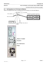
Detector Dose Measurement
Detector Dose measurements according to DIN 6868-152.
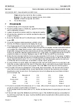
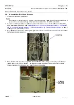
1. Remove the Bucky and install the dose measure support tool.
2. Place the Bucky and the 45 mm acrylic plate on the top of support tool.
3. Install the 6M Dose probe in the lower position.
4. Perform exposures in manual mode with parameters as close as possible to the AOP exposure
parameter (Table
) and measure the detector dose (1mR=8,76*10
-6
µGy).
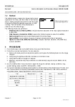
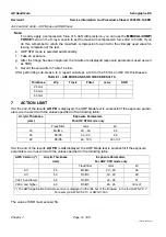
TABLE 3 -
The measured detector dose has to be less than 75 µGy for STD and Dose mode.
6-6
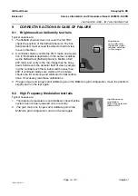
Contrast Mode Deactivate
Note:
Using the STD mode should satisfy most needs. However, if a higher priority is given to the dose
delivered to the patient, the DOSE mode may be selected instead. If a higher priority is given to
the contrast to noise ratio in images, the CNT mode may be selected. It is important to understand
that any improvement in contrast to noise ratio is done at the cost of an increase in glandular dose
and vice versa; a decrease in glandular dose will yield a reduction in contrast to noise ratio. For
more information on evaluating which priority to select, consult with your interpreting physician or
radiologist.
In case of site specific requirements, the access to CNT mode can be restricted as described in
Deactivate the Contrast Mode on page 789
7
POST-REQUISITES
On the X-ray Console, select
MEDICAL/DECOMP/DECOMP/YES
to activate the auto decompression
feature.
AOP mode
Spectrum (Target/Filter,
kVp)
mAs
Detector Dose, measured
by dosimeter, µGy
CNT
RhRh29
STD
RhRh29
Dose
RhRh29