GE Healthcare
Senographe DS
Revision 1
Service Information and Procedures Class A 2385072-16-8EN
Job Card ELE A037 - Image Acquisition and ACR Score
Page no. 705
Chapter 7
JC-ELE-A-037.fm
Job Card ELE A037 - Image Acquisition and ACR Score
Chapter 7
1
SUPPLIES
None
2
TOOLS
(16 cells Nuclear Associates 18-220 or RMI-156)
3
REQUIRED EFFORT
Personnel: 1 Field Engineer
Time: 8 minutes
4
SAFETY PRECAUTIONS.
The procedures in this Job Card produce X-rays. Ensure that you take appropriate precautions when
operating with X-rays (for example, stay behind the radiation screen during an X-ray exposure).
5
PREREQUISITES
The system must have been correctly installed and calibrated.
6
TEST DESCRIPTION
6-1
ACR Phantom
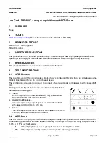
The phantom used in this procedure is a block of acrylic containing 16 cells. Each cell simulates an ana-
tomical structure which can be found in breast tissue.
The acrylic gives attenuation equivalent to a breast of average density compressed to a thickness of 45
mm.
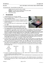
Starting from the top left-hand corner, as shown in the illustration,
the cells are of three types:
1. Fibers.
Six cells represent fibrous calcifications; they contain fibers
with sections from 1.56 mm to 0.40 mm.
2. Specks (Calcifications).
Five cells represent groups of specks or microcalcifications,
with sizes from 0.54 mm to 0.16 mm.
3. Masses.
Five cells represent tumors or masses, with sizes from 2 mm to
0.25 mm; each has a different attenuation.
6-2
ACR Score
The ACR Score check program obtains and displays an image of the phantom. After suitable adjustment
of the viewing parameters, the user notes the visibility of the object in each cell, using a 3-point scale of
1, 0.5, or 0. For each cell, score:
-
1
If the object (fiber, mass, or group of six specks) is completely visible.
-
0.5
If the object is partially visible, according to the following guidelines;


