Image Optimization 5-19
5.6
PW/CW Doppler Mode
PW (Pulsed Wave Doppler) mode or CW (Continuous Wave Doppler) mode is used to provide
blood flow velocity and direction utilizing a real-time spectrum display. The horizontal axis
represents time, while the vertical axis represents Doppler frequency shift.
PW mode provides a function for examining flow at one specific site for its velocity, direction and
features. CW mode proves to be much more sensitive to high-velocity flow display. Thus, a
combination of both modes will contribute to a much more accurate analysis.
CW imaging is an option.
5.6.1 Basic Procedures for PW/CW Mode Exam
1. Select a high-quality image during B mode or B + Color (Power) mode scanning, and adjust to
position the area of interest in the center of the image.
2. Tap [PW]/[CW] on the right side of the operating panel to enter PW/CW sampling line
adjustment status.
The sampling status will be displayed in the image parameter area in the top-left corner of the
screen as follows:
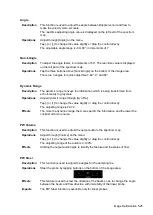
PW Sampling Line
Adjustment
SV
Angle
SVD
CW Sampling Line
Adjustment
Angle
CW Focus Depth
3. Set the position of the sample line by dragging the sampling line; drag the SV gate to place the
SV on the target.
4. Adjust the angle and SV size according to the actual situation: drag the PW angle line to
change the angle, pinch on the image area to adjust SV size.
5. Tap [PW]/[CW]/[Update] or double-click the sampling line to enter PW/CW mode and perform
the examination. You can also adjust the SV size, angle and depth in real-time scanning.
6. Adjust the image parameters during PW/CW mode scanning to obtain optimized images.
Tap [Image] to open the image menu. Adjust the parameters to optimize the image.
7. Perform other operations (e.g., measurement and calculation) if necessary.
Tap [Update] to switch between B (B+Color) and PW image.
5.6.2
PW/CW Mode Image Parameters
In PW/CW mode scanning, the image parameter area in the top-left corner of the screen displays
the real-time parameter values as follows:
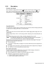
PW
Parameter
F
G
PRF
WF
SVD
SV
Angle
Meaning
Frequency
Gain
PRF
Wall
Filter
SV
Position
SV Size
Angle
CW
Parameter
F
G
PRF
WF
SVD
Angle
Meaning
Frequency
Gain
PRF
Wall Filter
SV Position
Angle
Summary of Contents for TE5
Page 1: ...TE7 TE5 Diagnostic Ultrasound System Operator s Manual Basic Volume ...
Page 2: ......
Page 6: ......
Page 12: ......
Page 24: ......
Page 36: ......
Page 54: ......
Page 110: ......
Page 115: ...Display Cine Review 6 5 6 Tap Return on the screen or tap Freeze to exit image compare ...
Page 120: ......
Page 124: ......
Page 156: ......
Page 174: ......
Page 192: ...12 18 Setup Select Advanced and do as follows Select MAPS and do as follows ...
Page 202: ...13 2 Probes and Biopsy C5 2s L12 4s L7 3s P4 2s L14 6s C11 3s L14 6Ns V11 3Ws P7 3Ts 7LT4s ...
Page 226: ...13 26 Probes and Biopsy NGB 034 NGB 035 ...
Page 250: ......
Page 272: ......
Page 276: ...A 4 Wireless LAN Tap Add Manually create a network profile to set ...
Page 282: ......
Page 318: ......
Page 322: ......
Page 323: ...P N 046 006959 07 1 0 ...


