ZEISS
Left Tool Area and Hardware Control Tools
ELYRA 7
196
000000-2262-999
03/2019 V_02
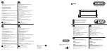
Expand the
Localizer
field to have access to the fit
model used to localize peaks (Fig. 319). Three fit
models are available that can be selected by the
Fit
Model
drop down menu (Fig. 320) if 2D is
selected from the Dimension drop down menu:
−
x,y 2D Gauss Fit
−
Mask fit
−
File PSF
If
3D
is selected from the
Dimension
drop down
menu, only
File PSF
is available. You should load
an Experimenatl PSF with linked look up table for
localization precision in order to obtain localization
precisions in z in addition to x and y.
If
x,y 2D Gauss Fit
is selected, the intensity
distributions of the identified peaks are fitted to a
full 2D Gaussian function. The fit in each direction
takes into account the influence of the other
direction (x,y 2D Gauss).
If
Mask Fit
is selected, a Gaussian mask with a
defined PSF width is used to match as best as
possible the intensity distribution of a peak in x
and y (Fig. 321). The PSF width can be set via the
PSF Width slider or input spin box. Default value is
the theoretical calculated PSF based on the
objective lens and laser line used.
Note that with the
Mask Fit
only one
predefined PSF width will be used.
Computation of the Fit will be faster for the
Mask Fit
, but less precise.
If
File PSF
is selected, you can select a PSF that will be used (Fig. 322). To this end press the
PSF file
button. This will open Windows Explorer, where you can select an approriate file, the name of which will
be displayed in the
PSF file
display box.
The fit model in 2D defines the PSF width, which will be used to fit to the experimental results. It
does not correspond to the size of the peak that is defined by the Peak Mask Size and that in the
end will define the overlapping degree of neighboring peaks. In 3D the PSF with the attached look
up table is a simulation where the PSF was modulated.
The settings of the
Localizer
will not influence the peak finding process and hence the preview
image.
In the Processing
SMLM
function, peak finding and localization are always linked. If you want to
just view localized peaks (molecules), you might select in the
SMLM-Render
tab of the SMLM
image in the
Display
drop down menu
Centroids
.
Fig. 319
Localizer Expansion field with x,y 2D
Gauss Fit selected
Fig. 320
Fit Model drop down menu
Fig. 321
Localizer Expansion field with Mask Fit
selected
Fig. 322
File PSF menu