5-4 Image Optimization
5.2
B Mode Image Optimization
B mode is the basic imaging mode that displays real-time images of anatomical tissues and
organs.
5.2.1 Basic Procedures for B Mode Imaging
1. Enter the patient information. Select an appropriate probe and exam mode.
2. Tap [B] on the right side of the operating panel to enter B mode.
3. Tap [Image] to open the image menu. Adjust the parameters to optimize the image.
4. Perform other operations (e.g., measurement and calculation) if necessary.
Tip: tap [B] to return to B mode at any time (except LVO mode).
5.2.2 B Mode Parameters
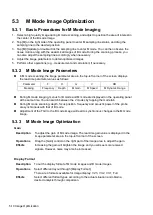
In B mode scanning, the image parameter area in the top-left corner of the screen displays the
real-time parameter values as follows:
Parameter
F
D
G
FR
DR
Meaning
Frequency Depth Gain Frame Rate Dynamic Range
5.2.3 B Mode Image Optimization
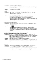
Frequency (Image Quality)
Description
To switch between the fundamental frequency and harmonic frequency as well
as select the corresponding frequency type. The real-time frequency value is
displayed in the image parameter area in the top-left corner of the screen, and if
harmonic frequency is used “F H” is displayed as the harmonic frequency value.
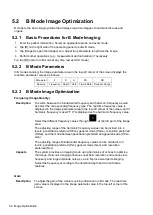
Select the different frequency values through
at the left part of the image
area.
The adjusting range of the harmonic frequency values can be divided into 4
levels: penetration preferred (HPen), general mode (HGen), resolution preferred
(HRes), and the mode between penetration preferred and general mode (HPen-
Gen).
The adjusting range of fundamental frequency values can be divided into 3
levels: penetration preferred (Pen), general mode (Gen), and resolution
preferred (Res).
Impacts
The system provides an imaging mode using harmonics of echoes to optimize
the image. Harmonic imaging enhances near-field resolution and reduces low-
frequency and large amplitude noise, so as to improve small parts imaging.
Select the frequency according to the detection depth and current tissue
features.
Gain
Description
To adjust the gain of the whole receiving information in B mode. The real-time
gain value is displayed in the image parameter area in the top-left corner of the
screen.
Содержание TE5
Страница 1: ...TE7 TE5 Diagnostic Ultrasound System Operator s Manual Basic Volume ...
Страница 2: ......
Страница 6: ......
Страница 12: ......
Страница 24: ......
Страница 36: ......
Страница 54: ......
Страница 56: ...4 2 Exam Preparation 4 1 1 New Patient Information The Patient Info screen is shown as follows 2 1 3 ...
Страница 110: ......
Страница 115: ...Display Cine Review 6 5 6 Tap Return on the screen or tap Freeze to exit image compare ...
Страница 120: ......
Страница 124: ......
Страница 156: ......
Страница 174: ......
Страница 192: ...12 18 Setup Select Advanced and do as follows Select MAPS and do as follows ...
Страница 202: ...13 2 Probes and Biopsy C5 2s L12 4s L7 3s P4 2s L14 6s C11 3s L14 6Ns V11 3Ws P7 3Ts 7LT4s ...
Страница 203: ...Probes and Biopsy 13 3 7L4s P10 4s L20 5s P7 3s L14 5sp SC6 1s SP5 1s 6CV1s L9 3s C5 1s L11 3VNs C4 1s ...
Страница 222: ...13 22 Probes and Biopsy No Name Description 8 Grooves of the needle guided bracket Matched with the tabs of the probe ...
Страница 226: ...13 26 Probes and Biopsy NGB 034 NGB 035 ...
Страница 250: ......
Страница 272: ......
Страница 276: ...A 4 Wireless LAN Tap Add Manually create a network profile to set ...
Страница 282: ......
Страница 318: ......
Страница 322: ......
Страница 323: ...P N 046 006959 07 1 0 ...

