Checks
6-8
6.4
Image Checks
6.4.1 B/W image phantom data checks and image records
6.4.1.1
System setups
The user-defined setups are adopted for all the setups which aren’t mentioned in this manual.
For the setups changed due to special reasons, they shall be recorded as added information.
6.4.1.2
Image records and archives
The printed images shall be archived with the recorded data.
6.4.1.3
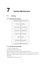
Flowchart for checks
Check lateral and axial resolutions
↓
Check penetration
↓
Check spot characteristics
↓
Record and check images
Perform the above checks for all transducers used.
6.4.2 Check phantom data
6.4.2.1
Lateral/axial resolutions
1. Smear ultrasound gel on the phantom, and use the transducer to examine.
2. After a good-quality image is obtained, freeze and record the image.
Condition: system preset parameters
6.4.2.2
Penetration
1)
Smear ultrasound gel on the phantom, and use the transducer to examine.
2)
Adjust GAIN to make the spots of soft tissue displayed in the deepest position.
3)
Measure the noise and the depth of the soft tissue boundary, and record the image
for measuring the depth.
6.4.2.3
Spot characteristics
Evaluate the images for quality after the system has been used for a long period of time,
including gain and periodically recorded images.
Summary of Contents for DC-6
Page 1: ...DC 6 DC 6T DC 6Vet Diagnostic Ultrasound System Service Manual...
Page 2: ......
Page 20: ...2 1 2 System Overview 2 1 System Appearance 2 1 1 Complete System with CRT Monitor...
Page 23: ...System Overview 2 4 2 2 LCD Monitor...
Page 26: ...System Overview 2 7 2 2 3 Lever of upper support arm...
Page 66: ...4 1 4 System Structure and Assembly Disassembly 4 1 Exploded View of Complete System...
Page 101: ...System Structure and Assembly Disassembly 4 36 Power boards Card detacher...
Page 191: ...P N 2105 20 40473 V10 0...


