GE Healthcare
Senographe 2000 D Acquisition System
REV 1
OM 5179217–1–100
129
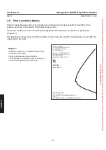
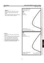
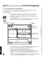
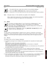
Model 2:
Patient data at top, followed by view data.
Institution, image and exposure data at
bottom.
Top and bottom annotation areas are aligned
to the border opposite the chest wall.
Model 3:
View data at top, followed by exposure date
and time.
Patient data at bottom left, institution data at
bottom right, followed by exposure data.
The top annotation area is aligned to the
border opposite the chest wall. All bottom
annotations are restricted to the image footer
so as to avoid overlap with the image.
CHAP
. 1
1
FOR
TRAINING
PURPOSES
ONLY!
NOTE:
Once
downloaded,
this
document
is
UNCONTROLLED,
and
therefore
may
not
be
the
latest
revision.
Always
confirm
revision
status
against
a
validated
source
(ie
CDL).

