


136
Basic User Manual
10 4D Imaging
10
�
1 Acquiring 4D Images
The following description uses 4D imaging performed with the VC6-2 probe as an
example.
10
�
1
�
1 Entering the Real-Time 4D Imaging
Perform the following steps as follows.
1.
Enter the patient information, select the VC6-2 probe and an exam type (such as the
obstetric exam used throughout this chapter) to enter the B mode.
2.
Optimize imaging parameters in the B mode.
3.
Apply adequate gel on the patient.
4.
Acquire a high quality B-mode image.
5.
Tap
3D/4D
on the touch screen to enter the inactivated 4D mode by default.
NOTE:
When using a volume probe, you can set the default inactivated 3D or 4D mode.
For details, refer to Section 4.1.4 Defined-Key Settings.
The 2D imaging with a ROI and a sample line is displayed in the inactivated 4D
mode, only the data in the ROI is acquired for 4D imaging.
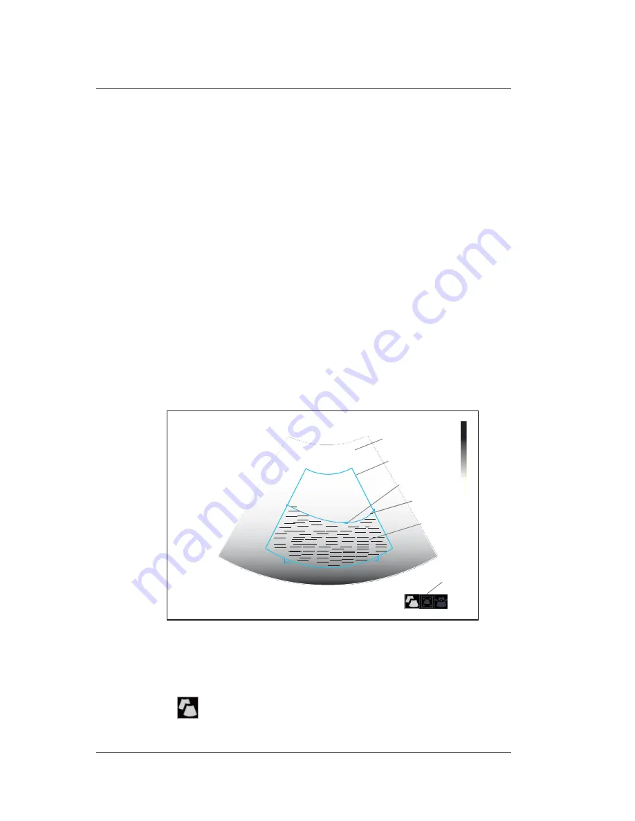
Figure 10-1 Inactivated 4D Imaging Screen
6.
Adjust the ROI and sample line.
You can press the confirm key to select the desired status icon.
−
If
is highlighted, use the trackball to position the ROI.
Status Icon
Sample Volume
D
16.5cm
Anchor
Angle
55
Sample Line
Quality H
ROI
PWR 70
FRQ 3-4.8
2D Imaging
47
3/1
255
3/30
FPS
D/G
GN
I/P
Summary of Contents for P25 EXP
Page 1: ...User Manual P25 EXP Ultrasound System Version 1 4...
Page 5: ......
Page 12: ...Contents VIII Basic User Manual D 3 Disinfectant 200 AppendixEAcousticOutputData 201...
Page 48: ...This page is intentionally left blank...
Page 168: ...This page is intentionally left blank...
Page 176: ...This page is intentionally left blank...
Page 200: ...This page is intentionally left blank...
Page 213: ...201 Appendix E Acoustic Output Data Please consult the accompanying CD...


