Indicates the chosen display map for the image. The display map can be changed
using the display map control.
Image area
This is the area where the image data acquired by the transducer appears. This is the
area of the window that the system exports—along with header information—when
you export a stored image and configure your export to send only the image area.
The image area is not interactive, you control the content of the image area from the
control panel.
This area is also known as the workspace.
Transducer orientation indicator
The blue dot corresponds to the orientation ridge on the transducer nose and indicates
the orientation relative to the anatomy.
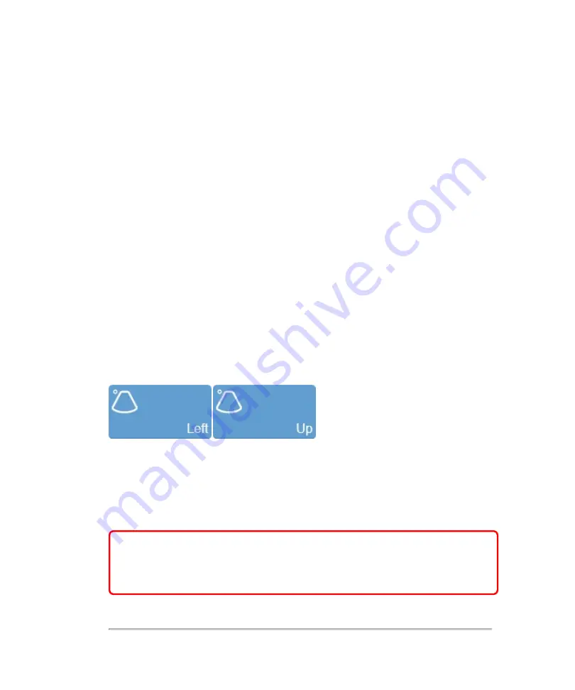
Tap either of the orientation buttons to flip the image between the following options:
Right
,
Left
,
Up
and
Down
.
Image scale
Indicates, in mm, the distance from the face of the transducer to the tissue being
imaged.
WARNING:
The Vevo MD Imaging System uses ultra high frequency (UHF)
series transducers. Each UHF transducer model has a different image scale
that does not start at 0.0 mm, but includes an offset. Please be aware of this
when placing measurements.
200
Scanning
Summary of Contents for VisualSonics Vevo MD
Page 1: ......
Page 2: ......
Page 12: ...12 ...
Page 69: ...System settings 69 ...
Page 70: ...70 System settings ...
Page 77: ...3 Tap DICOM Setup Connectivity 77 ...
Page 146: ...2 Tap User Management in the list on the left 146 System settings ...
Page 168: ...Review images screen 1 Next and previous image 2 Scan 3 Export 4 Delete 168 Patient ...
Page 461: ...zoom while scanning 2D control 226 Color Doppler Mode control 265 Index 461 ...
Page 462: ...462 Index ...
Page 463: ...51370 01 1 0 51370 01 ...


