APPLIED OPERATIONS
/Image Acquisition
OPERATION INSTRUCTIONS
2 - 9
Page
2-2-1-1 Configuring the Microscope
Set the light path so that the image can be observed through the microscope.
1. Display the [Acquire] panel.
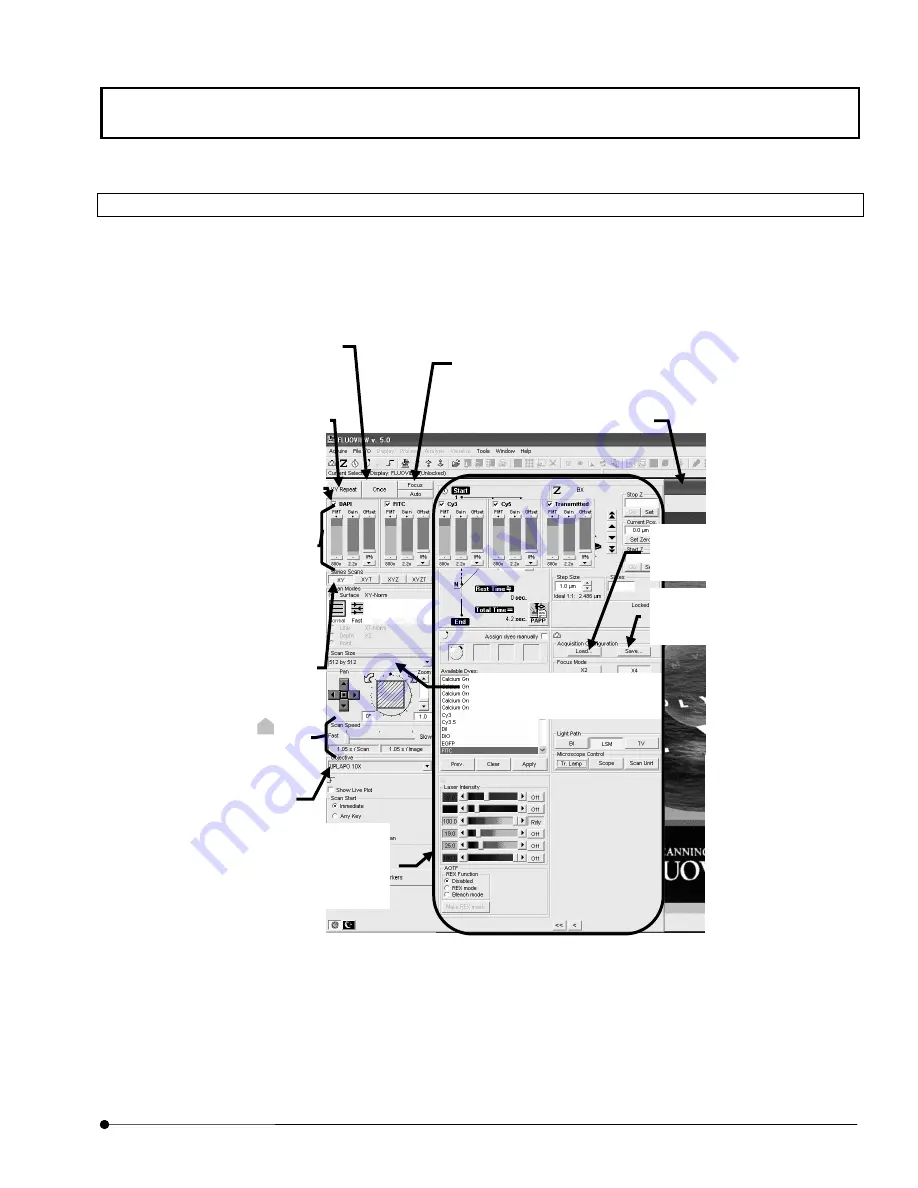
Fig. 2-1 [Acquire] Panel
<Focus> button
The repetition images can be acquired at a high speed.
Use this button to find an optimum image.
<Once> button
Acquires an image in the currently
selected observation mode and display
the image in the [Live] panel.
[Settings], [Z Stage], [Time Series],
[Dyes] and [Lasers] sub-panels
These panels are used to set the
information required for image
acquisition.
<XY Repeat> button
Repeats XY scanning to display images
successively in the [Live] panel.
Option buttons
Check a button to select one of the
displayed observation modes.
[Scan Speed] group box
Sets the scan speed. Sliding
to the
left of the scale increases the scan
speed and to the right decreases it.
[PMT], [Gain] and [Offset] LED
sliders
These can be adjusted independently.
Increasing the PMT voltage improves
the sensitivity.
Increasing the Gain brightens the
image.
Increasing the Offset darkens the
image.
[Ch1], [Ch2] and [Ch3] check boxes
Check to select the image acquisition
channel.
Select the magnification of the
objective on the microscope.
The acquired image is displayed.
[Scan Size] drop down list
Select the image acquisition size.
<Load> button
Loads the observation conditions
at image acquisition.
<Save> button
Saves the observation conditions
at image acquisition.
Содержание Fluoview FV1000
Страница 2: ......
Страница 12: ......
Страница 22: ......
Страница 356: ......
Страница 397: ...APPLIED OPERATIONS Viewing 3D Image OPERATION INSTRUCTIONS 2 3 1 3 Page Fig 2 130 Panel Showing Stereo 3D Images...
Страница 446: ......
Страница 452: ......
Страница 464: ......
Страница 476: ......
Страница 482: ......
Страница 484: ......
Страница 486: ......
Страница 524: ......
Страница 534: ......
Страница 536: ......
Страница 539: ......


