GE
V
OLUSON
i / V
OLUSON
e
D
IRECTION
KTI106052, R
EVISION
10
S
ERVICE
M
ANUAL
Chapter 4 - Functional Checks
4-7
8
ß-VIEW
BT
Version:
BT-Version:
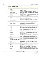
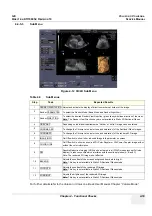
This function allows the adjustment of the Volume O-Axis position of 3D
probes in 2D Mode. The green line in the displayed symbol indicates the
position of the acoustic block.
This feature is only available at systems with
BT´07
software and higher.
9
SRI LOW / SRI HIGH
(Speckle Reduction Imaging)
Speckle Reduction Imaging is a smoothing type filter to reduce speckle in the
ultrasound image.
10
2D+2D/XXX
Changes the Single image display to two simultaneous half images. The left
frame shows only the 2D Mode image. The right frame shows the 2D Mode
image with xxx (xxx = SRI or CRI) information.
11
FFC (Focus and Frequency Composite)
FFC combines a low frequency to increase the penetration and higher
frequency to keep a high resolution.
It reduces speckle and artifacts in the 2D image.
12
LINEAR / TRAPEZOID
Advantage of the Trapezoid Mode: The scan area is very increased in
relation to the linear display by steering the ultrasound lines in the border of
the probe.
13
High Resolution Zoom
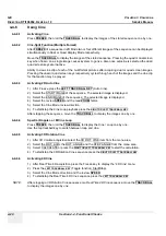
Press the
MAGNIFY GLASS
key (nearby the trackball) in write mode. The
displayed zoom box can be placed over the entire 2D image area, also the
size and position of the zoom box can be changed. Press the
MAGNIFY GLASS
key again to activate the zoom and again to exit the High
Resolution Zoom function.
14
Harmonic Imaging
Press the
HI
key on the control panel to switch on/off the Coded Harmonic
Imaging function in 2D Mode provided the active probe allows this function.
15
ANGLE
Press the Soft-menu buttons to select a part of interest of the 2D image.
The advantage of the decreased field-of-view is an increased 2D frame rate
due to the smaller sector width.
16
FOC DEPTH
Press the Soft-menu buttons to select the depth position of the actual focus
zone(s). Arrows at the left edge of the 2D image mark the active focal
zone(s) by their depth position.
17
FOC NUM:
Pressing the Soft-menu buttons increases the number of transmit focal zone,
so that you can tighten up the beam for a specific area.
18
ZOOM
Press the Soft-menu buttons for Image magnification (Pan Zoom) in read
and write mode.
19
FREQ: Resol.
HARM.FREQ. in case of Harm. Imaging
Press the Soft-menu buttons to adjust the range of the receive frequency;
high resolution/lower penetration, mid resolution/mid penetration, or lower
resolution/high penetration
20
QUALITY
Control to improve the resolution by reducing the frame rate.
Respectively reducing the resolution by increasing the image frame rate.
21
OTI (Otimized Tissue Imaging)
OTI™ allows to “fine tune” the system for scanning different kinds of tissue.
22
PERSIST
Persistence is a temporal filter that averages frames together.
This has the effect of presenting a smoother, softer image.
23
ENHANCE
Enhance brings out subtle tissue differences and boundaries by enhancing
the gray scale differences corresponding to the edges of structures.
Adjustments to M Mode's edge enhancement affects the M Mode only.
24
REJECT
Selects a level below which echoes will not be amplified (an echo must have
a certain minimum amplitude before it will be processed).
Table 4-3
2D Mode Functions
Step
Task
Expected Results
Содержание Voluson i BT06
Страница 2: ......
Страница 11: ...GE VOLUSON i VOLUSON e DIRECTION KTI106052 REVISION 10 SERVICE MANUAL ix ZH CN KO...
Страница 44: ...GE VOLUSON i VOLUSON e DIRECTION KTI106052 REVISION 10 SERVICE MANUAL xlii Table of Contents...
Страница 514: ...GE VOLUSON i VOLUSON e DIRECTION KTI106052 REVISION 10 SERVICE MANUAL IV Index...
Страница 515: ......

