5-28 Image Optimization
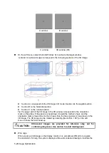
On the soft menu, select [Current Window: X] to activate the target window.
A, B and C sectional images correspond to the following sections of the 3D image.
Section A: corresponds to the 2D image in B mode. Section A is the sagittal section .
Section B: is the horizontal section.
Section C: is the coronal section.
Tip: the upper part of the 3D image in the 3D window corresponds to the orientation
mark on the probe. If the posture is he
ad down (toward the mother’s feet), and the
orientation mark is toward the mother’s head, then the fetus posture is head down in the
3D image. The 3D image can be rotated by selecting [Quick Rot.: 180°] on the soft
menu to show the fetus head-up.
CAUTION:
Ultrasound images are provided for reference only, not for
confirming diagnoses. Use caution to avoid misdiagnosis.
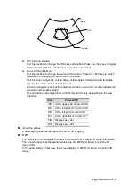
Wire cage
When viewing a 3D image on the display monitor, it is sometimes difficult to recognize
the orientation. To help, the system displays a three-dimensional drawing to illustrate the
A window
B window
C window
3D window (VR)
Содержание DP-50 Exp Vet
Страница 2: ......
Страница 34: ...2 6 System Overview 2 6 Introduction of Each Unit Right View Left View...
Страница 42: ......
Страница 68: ......
Страница 128: ......
Страница 148: ......
Страница 166: ...10 18 DICOM For details on tast manager see 9 6 Animal Task Manager...
Страница 180: ......
Страница 220: ......
Страница 224: ......
Страница 236: ......
Страница 242: ......
Страница 248: ......
Страница 249: ...Acoustic Output Reporting Table 60601 2 37 C 1 Appendix C Acoustic Output Reporting Table 60601 2 37...
Страница 342: ...D 2 Printer Adapter Type Model SONY X898MD...
Страница 343: ...P N 046 017713 02 1 0...


