16 Probes and Biopsy
Operator’s Manual
16 - 19
needle-guided bracket, immediately stop using it and contact MINDRAY
Customer Service Department or sales representative.
•
Disposable brackets are packaged sterile and are single-use only, and the
method of sterilization is irradiation. Do not use if the sterile packaging is
open or broken, and do not reuse or resterilize the disposable brackets.
•
DO NOT use a needle-guided bracket when scanning is performed. The
needle may advance in an incorrect direction and possibly injure the
patient.
Never perform a biopsy during image scanning.
•
DO NOT freeze an image while performing biopsy procedure.
•
During biopsy procedures, the needle may deviate from the desired course
due to the tissue characteristics or the type of needle. In particular, needles
of small diameters may deviate to a greater degree.
•
Disinfect the probe and sterilize needle-guided bracket before and after
each ultrasound-guided biopsy procedure is performed. Fail to do so may
cause the probe and the needle-guided bracket become sources of
infection.
•
The needle mark displayed on the ultrasound image does not indicate the
actual position of the biopsy needle. Therefore, it should only be used as a
reference. Always monitor the relative positions of the biopsy needle during
the procedures.
•
Adjust the needle mark before the biopsy procedure is performed.
•
When performing biopsy procedures, use only sterile ultrasound gel that is
certified to be safe. And manage the ultrasound gel properly to ensure that
it does not become a source of infection.
•
When performing the operation concerning biopsy, wear sterile gloves.
•
Image of the biopsy target and the actual position of the biopsy needle:
Diagnostic ultrasound systems produce tomographic plane images with
information of a certain thickness in the thickness direction of the probe.
(That is to say, the information shown in the images consist all the
information scanned in the thickness direction of the probe.) So, even
though the biopsy needle appears to have penetrated the target object in
the image, it may not actually have done so. When the target for biopsy is
small, dispersion of the ultrasound beam may lead to image deviate from
the actual position. Pay attention to this.
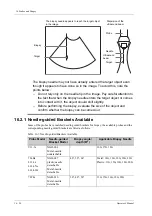
If the target object and the biopsy needle appear in the image as shown in
the figures below (For reference only):
Содержание Ana
Страница 2: ......
Страница 50: ...This page intentionally left blank...
Страница 60: ...This page intentionally left blank...
Страница 110: ...This page intentionally left blank...
Страница 116: ...This page intentionally left blank...
Страница 166: ...This page intentionally left blank...
Страница 176: ...This page intentionally left blank...
Страница 194: ...This page intentionally left blank...
Страница 220: ...This page intentionally left blank...
Страница 288: ...This page intentionally left blank...
Страница 304: ...This page intentionally left blank...
Страница 308: ...This page intentionally left blank...
Страница 316: ...This page intentionally left blank...
Страница 337: ......
Страница 338: ...P N 046 018835 00 2 0...


