background image

13  Probes and Biopsy 

122 

Basic User Manual

Orientation 

mark

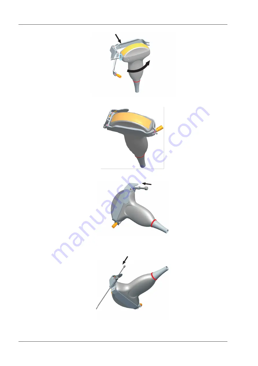
6.  Attach the biopsy bracket to the probe and fasten the biopsy bracket with the locking screw.

7.  Press the handle and insert the biopsy bracket tube into the biopsy bracket.

8.  Insert the biopsy needle into the guide tube and make sure that the biopsy bracket is firmly attached to the 

probe.

9.  Unfold another probe sheath, and apply an appropriate amount of coupling gel to the inside of the sheath. 

Содержание EVUS 8

Страница 1: ...C d Rev 02 77000001436 EVUS 8 OWNER S MANUAL English...

Страница 2: ...nd Diagnostic Equipment Model Evus 8 Brand Saevo Respons vel T cnico Daniel R de Camargo CREA SP 5062199650 ANVISA Registration n 10069210070 Alliage S A Industrias M dico Odontol gico Rodovia Abr o A...

Страница 3: ...ity and performance of this product only if all the following requirements are met All installation operations expansions changes modifications and repairs of this product are conducted by the manufac...

Страница 4: ...Item Meaning Click Move the cursor to the controls on the display and press the confirm key on the control panel Select a menu item or a key following the path...

Страница 5: ...10 2 3 2 Side View 10 2 3 3 Rear Panel 11 2 3 4 Battery Compartment 11 2 3 5 Control Panel 12 2 3 6 Basic Screen 15 3 Preparing the System 17 3 1 Positioning the System 17 3 2 Powering the System 17...

Страница 6: ...m 47 5 1 Acquiring Patient Information 47 5 1 1 Beginning a New Patient 47 5 1 2 Retrieving Archived Information 49 5 1 3 Using DICOM Worklist 50 5 2 Pausing Resuming an Exam 50 5 3 Completing Discont...

Страница 7: ...otating Magnifying Images 87 8 2 7 Optimizing 3D Image 88 8 2 8 Observing reviews by the slice 88 8 2 9 Setting the Scan Mode 89 8 2 10 Adjusting Z Angle Z Scale 89 8 2 11 Restoring the Image 90 8 2 1...

Страница 8: ...12 Working with DICOM 107 12 1 Verifying Connectivity 107 12 2 DICOM Storage 107 12 3 DICOM Print 108 12 4 DICOM Worklist 108 12 5 MPPS 109 12 6 Storage Commitment 110 12 7 DICOM Queue 111 13 Probes...

Страница 9: ...in Case of Destruction of the System 129 14 7 Customer Service 130 Appendix A Specifications 131 Appendix B EMC Guidance and Manufacturer s Declaration 132 Appendix C In Situ Derated and Water Value I...

Страница 10: ...This page is intentionally left blank...

Страница 11: ...red in the use of any application Otherwise it may result in system damage or serious injury 1 2 Safety Precautions Read and understand all precautions in this manual before attempting to use the syst...

Страница 12: ...efore powering it on Ensure that the temperature and humidity inside and around the system are equivalent before an operation Do not use the system around a strong electric field a strong electromagne...

Страница 13: ...robe from the system after freezing an image or powering off the system Otherwise the system or the probe could be damaged 1 2 4 Biohazard Considerations To minimize the risk of cross contamination or...

Страница 14: ...oeffects Considerations for the Safety of Diagnostic Ultrasound J Ultrasound Med Sept 1988 Vol 7 No 9 Supplement sometimes referred to as the StoweReport which reviewed available data on possible effe...

Страница 15: ...the limit The surface temperature of the probe is displayed on the lower left side of the screen PAT 37 TIP 28 Patient Threshold Temperature Transducer Tip Temperature 1 3 5 Imaging Functions that Ch...

Страница 16: ...Direct current Degree of IP protection Non ionizing electromagnetic radiation Manufacturer Date of manufacture Follow instructions for use Network port USB port Fragile AUDIO output Keep dry Maximum s...

Страница 17: ...only Rechargeable for battery only This symbol indicates that waste electrical and electronic equipment must not be disposed of as unsorted municipal waste and must be collected separately Please cont...

Страница 18: ...This page is intentionally left blank...

Страница 19: ...mponents the control panel the key panel and the basic screen 2 1 System Configuration The standard system configuration mainly consists of the followings items Main unit Probes Others See the Packing...

Страница 20: ...l 1 2 3 4 Figure 2 2 Front View 1 LCD Monitor 2 Probe Holder 3 Control Panel 4 Release Latch 2 3 2 Side View 1 2 3 4 5 6 Figure 2 3 Side View 1 Audio Output Port 2 S VIDEO Port 3 Network Port DICOM 4...

Страница 21: ...Panel 1 Rating Label 2 Video Printer Port 3 Engineering USB Port 4 Grounding Terminal 5 Foot switch Port 6 BNC Video Output Port 7 ECG Port 8 AC Adapter Port 9 Probe Port 10 Probe Lock 2 3 4 Battery C...

Страница 22: ...Description Patient Select it to enter the New Patient screen Report Select it to enter the Report screen File Select it to display the Patient Exam List screen Or select it to enter the View Image s...

Страница 23: ...ect it to reverse the image left or right Up Down Select it to reverse the image up or down DEPTH Use it to adjust the scan depth Flip it downwards or upwards to adjust the depth FOCUS Use it to adjus...

Страница 24: ...k symbols from the display Annot Select it to annotate an image with typed words Distance Select it to activate the basic measurement and calculation functions Select it to perform a distance measurem...

Страница 25: ...atus Indicator Indicator Description Indicates the battery discharging status Indicates the battery charging status Indicates the connecting status of the mains supply 2 3 6 Basic Screen 0 5 10 15 9 c...

Страница 26: ...9 Operator 10 Measurement Result Area 11 Color Map in C PDI mode Grayscale Map Right 12 Depth Scale and Focal Position 13 Cine Loop Icon in frozen mode 14 Network Connection Status 15 Image Parameter...

Страница 27: ...AC adapter can only be used for power supply or battery charge of the system Other usages may damage the adapter or even cause an electric shock Ensure that the adapter is not damaged before use If d...

Страница 28: ...Do not expose the battery under the sunlight or leave it in the environment over 60 C Keep the battery away from fire and other heat sources during use and charge Do not pierce the battery with a shar...

Страница 29: ...ly 3 2 4 Status Indicator The indications of each status indicator are shown in Table 3 1 Table 3 1 Status Indicator Status Indicator Mains Supply Battery Battery Charging Indicator Yellow charging Of...

Страница 30: ...r off the system Press the button on the left side of the system to power off the system System standby If the ultrasound system is idle for a set period the system enters the standby mode automatical...

Страница 31: ...Otherwise the ultrasound system or the foot switch may be malfunctioning Connect the foot switch to the foot switch port of the ultrasound system as the figure shows below Used for image freezing Used...

Страница 32: ...SB drive 2 Connect the USB drive to the ultrasound system and then start up the ultrasound system 3 Select the Menu key to enter the System Setting menu and click Peripheral 4 Tick Static IP and input...

Страница 33: ...he available probe and the application mode screen as shown in Figure 4 1 XXX Figure 4 1 LCD Monitor Select the Menu key on the control panel to enter the System Setting menu To make settings on the S...

Страница 34: ...Setting KeyConfig Time Format Figure 4 2 General Settings Menu General Tab Item Description Hospital Name Enter the institution s name Freeze Response Select the feature to be enabled after you select...

Страница 35: ...nes Set it to Gun one biopsy guideline is displayed when performing the biopsy Set it to Needle two parallel biopsy guidelines are displayed when performing the biopsy These guidelines make up the gui...

Страница 36: ...following steps to perform a demonstration 1 Click Show Gallery to enter the Show Gallery screen as shown in Figure 4 4 Show Gallery Gallery Config Top Up Down Bottom Import Delete Load Default Exit F...

Страница 37: ...disable the one key save function Set it to ON the image or cine is saved automatically by selecting the Save key on the control panel in the frozen mode Set it to OFF you need to manually save the i...

Страница 38: ...ed cine Volume Tick it and select the U1 or U2 key to save the desired volume data Print Tick it and select the Save U1 or U2 key to save the desired image and print it out Send to USB Tick it and sel...

Страница 39: ...of the ultrasound system NetMask Set the netmask address of the ultrasound system Default Gateway Set the gateway address of the ultrasound system Speed Duplex Click Advance Speed and Duplex textboxe...

Страница 40: ...e network printer Add Printer Click the button and follow the on screen instructions to install a printer driver 4 3 Defining Annotation Library You can add modify delete or arrange the predefined ann...

Страница 41: ...own list beside Application of Available 3 Add desired body marks in the selected body mark library to the Selected list Select a body mark and then click to add it to the Selected list Click to add a...

Страница 42: ...system calculates BSA value automatically after you enter the height and weight of the patient on the Patient Information screen Cross Cursor Size Set the size of the cross cursor Measure Line Size S...

Страница 43: ...Measure DICOM Load Default About Exit Save Exit Bodymark Report No Growth Dev Display Figure 4 11 Measurement Settings Menu Application Tab Auto Man Trace After selecting an exam type from the applic...

Страница 44: ...by EFW Set the formula to estimate GA by EFW If it is set to None the system will not estimate GA by EFW and no corresponding values are displayed on the measurement report EFW GP SD Set the formula...

Страница 45: ...4 12 GA Table Growth Table Measurement Age w d 2SD mm Mean mm 2SD mm Unit Author Deviation 2SD 2SD w d Meas 1 2 3 4 5 6 7 8 9 10 11 12 13 14 15 16 17 18 19 2SD Mean 2SD mm mm mm Exit Save Exit Figure...

Страница 46: ...steps as follows 1 Select an exam type from the drop down list beside Application 2 Select Distance or Calc 3 Select an imaging mode in the List frame and click Collection or Measurement 4 Click Add...

Страница 47: ...ort logo 170 60 pixels bmp format to the ultrasound system Operation LOGO is highlighted after the USB drive is successfully detected by the ultrasound system Click Operation LOGO and then click the d...

Страница 48: ...Default About Exit Save Exit Bodymark Report Figure 4 16 DICOM Settings Menu Storage Service Tab Item Description Service List Select Append from the drop down list beside Service List input the rele...

Страница 49: ...server and adjust the relevant settings on the Commitment Storage tab Delete Add DICOM Storage Commitment System Setting Store C Store Worklist Print MPPS Service List StoreCommitment Remote HostName...

Страница 50: ...ment storage server after you input the settings for the Remote HostName IP address DICOM AE Title Port Number A summary displays if the connection is verified 4 7 3 DICOM Worklist Settings You can co...

Страница 51: ...e command Max Result Set the maximum number of patient records that can be sent to the ultrasound system Click Echo to verify the connection between the ultrasound system and the DICOM worklist after...

Страница 52: ...t the port number of the MPPS server Connect Timeout sec Set the time limit for the network connection timeout Repeat Count Set the number of repeated attempts for connecting to the MPPS server Dimse...

Страница 53: ...Service Tab Item Description Service List Select Append from the drop down list beside Service List input the relevant parameters information and click Add to add a new server Select any server from...

Страница 54: ...al image quality settings Copies Set the number of copies to print Film Session Label Enter a name to be applied on a group of the film labels Smoothing Type Set the value of magnification interpolati...

Страница 55: ...ding patient database Please use it wisely New Export Import Load Export Log Peripheral General Comment Measure DICOM Load Default About Exit Save Exit Bodymark Report Figure 4 22 Export Import Menu E...

Страница 56: ...acoustic output data of probes in different modes on the Acoustic Output tab System Setting General Acoustic Output Register Type Probe Type Exam Type Peripheral General Comment Measure Bodymark Repo...

Страница 57: ...panel to display the New Patient screen in Figure 5 1 New Patient Base Information Exam Information Patient ID Last Name First Name DOB M I Age D MM DD YYYY Sex Comments Accession Description Chief Co...

Страница 58: ...ea after you enter the weight and height You can select the calculation formula from the corresponding drop down list BP Enter the blood pressure for the current patient Comments Make comments for the...

Страница 59: ...To 05 04 2014 05 05 2014 Current Exam New Patient DICOM Queue PPS Screen Import Export Import Screen Export Screen Exit Exam Type SMP 05 05 2014 16 04 03 40 82M Exam Date Time Image Size HD Search Re...

Страница 60: ...OM Worklist You can acquire the patient information using the DICOM worklist if the ultrasound system is configured with the DICOM module For details refer to Section 12 4 DICOM Worklist 5 2 Pausing R...

Страница 61: ...tient to enter the New Patient screen Input the new patient information and click OK A dialogue box pops up as follows PPS Assisted Acquisition Protocol Setting Code Meaning Designator Reason Complete...

Страница 62: ...This page is intentionally left blank...

Страница 63: ...tent with the connected probe If a problem is found check that the probe is connected to the system firmly If the problem still exists please stop using the system immediately and contact the manufact...

Страница 64: ...ion preset in the Exam Name textbox 6 Select the department and exam type from the Exam Type and Exam Icon lists 7 Click Create Exam to create the customized application preset If the name of the appl...

Страница 65: ...Yes in the pop up dialogue box to restore factory defaults 6 2 Acquiring B Mode Images B mode imaging is intended to provide information of anatomical structure of soft tissue 6 2 1 Entering B Mode Th...

Страница 66: ...C Move the desired TGC slider left to decrease the value Move the desired TGC slider right to increase the value NOTE Modifications of TGC values cannot be saved if you shift to use another imaging mo...

Страница 67: ...pops up on the screen 2 Select Chroma by using the trackball or the or key 3 Rotate the Menu key or select the or key to choose a color scheme Compound Imaging Compound imaging is used to acquire a se...

Страница 68: ...optimization To adjust the adaptive image fusion 1 Select the Menu key in the real time scan A menu pops up on the screen 2 Select Adapt Fusion by using the trackball or the or key 3 Rotate the Menu...

Страница 69: ...nformation This item is applicable in transvaginal and transrectum scan To rotate the image 1 Select the Menu key in the real time scan A menu pops up on the screen 2 Select Image Rotate by using the...

Страница 70: ...e image Perform the following steps to acquire C mode images 1 Optimize a B mode image 2 Select the C key on the control panel to enter the C mode 0 5 10 15 9 9 cm s 86 2D Imaging Color Flow ROI 1 0 P...

Страница 71: ...r flow ROI Move the trackball to position color flow ROI Select the Set key to adjust the size of color flow ROI Select the Set key to reposition color flow ROI 4 Optimize the PDI mode image For detai...

Страница 72: ...f the LCD monitor as shown in Figure 6 6 you can use them to optimize C PDI TDI mode images You can adjust other parameters and display modes on the pop up menu by selecting the Menu key NOTE You can...

Страница 73: ...he position of zero velocity or frequency The velocity range in one direction can be increased or decreased by adjusting the baseline Baseline is used to eliminate the alias in the color flow imaging...

Страница 74: ...e amount of ultrasound acoustic power produced by the probe The adjustment range of the power is 0 100 and 10 can be adjusted each time The real time value of the power is displayed in the imaging inf...

Страница 75: ...nterest and then learn about the tissue motion along that line in M mode trace In M mode y axis is used to display the position or depth of the tissue and x axis is the time scale 6 4 1 M Mode The M l...

Страница 76: ...ased array probes when performing cardiac exams or convex probes when performing abdominal exams In the anatomical M mode the M line can be positioned perpendicular to the anatomical structure and be...

Страница 77: ...st all parameters in the real time M mode but can only adjust Chroma and Dis Format in the frozen M mode Gain M gain controls the overall brightness of the M trace To adjust the gain Rotate the B knob...

Страница 78: ...ble the video invert 1 Select Video Invert by pressing the corresponding function key 2 Rotate it to enable or disable the feature 6 5 Acquiring Spectral Doppler Images Spectral Doppler imaging is int...

Страница 79: ...all left or right 4 Adjust the sample volume gate Adjust the size of the sample volume gate by moving the trackball Rotate the Angle knob to increase or decrease the angle of the flow cursor 5 Select...

Страница 80: ...ived ultrasound energy to generate a spectral display CW Doppler is used to measure a high velocity the highest velocity at a specific site but no aliasing is created Perform the following steps to ac...

Страница 81: ...gure 6 15 CW Mode Imaging Screen X axis is the time scale Y axis is Doppler frequency scale including positive and negative indicators Select the Audio or Audio key to adjust the audio volume NOTE Onc...

Страница 82: ...a Video Invert and Dis Format in the frozen PW CW mode Gain PW CW gain is used to adjust the gain of the spectrum map Increasing the gain will brighten the image allowing you to see more received sign...

Страница 83: ...Baseline by using the trackball or the or key 3 Rotate the Menu key or select the or key to move the baseline towards the positive or negative direction 4 The speed scale on the y axis at the bottom r...

Страница 84: ...ts in the grayscale It is useful for optimizing the ultrasound image texture To adjust the dynamic range 1 Select DYN by pressing the corresponding function key 2 Rotate it to adjust the value Display...

Страница 85: ...modes To enable or disable the feature Select Simult in the activated PW mode or in the inactivated B PW mode 2 the two real time scans in the 2D and the PW modes are displayed NOTE Simult is only ava...

Страница 86: ...6 2 B C PDI TDI PW Perform the following steps to acquire the image 1 Optimize a C PDI TDI mode image 2 Select the PW key to enter the inactivated C PDI TDI PW mode 1 0 5 Spectral Doppler Line Sample...

Страница 87: ...to adjust the audio volume 6 Optimize the PW mode image For details refer to Section 6 5 3 Optimizing Spectral Doppler Images If needed select the Update key on the control panel to enter the inactiva...

Страница 88: ...This page is intentionally left blank...

Страница 89: ...e Elastic key on the key panel to enter the elastography imaging As Figure 7 1 shows the elastography image is displayed on the left and a B mode image in a real time B mode image is displayed on the...

Страница 90: ...lastography Images 7 Select the Elastic key again to exit 7 2 Optimizing Elastography Images After entering the elastography imaging the imaging parameters display at the bottom of the LCD monitor you...

Страница 91: ...ues and higher persistence values for smoother appearance To adjust the persistence 1 Select the Menu key in the real time scan A menu pops up on the screen 2 Select Persist by using the trackball or...

Страница 92: ...This page is intentionally left blank...

Страница 93: ...the B mode 3 Apply adequate gel on the patient 4 Acquire a B mode image of high quality 5 Select the 3D 4D key on the control panel to enter the inactivated 3D mode NOTE You should select the Freeze...

Страница 94: ...ging Screen As the above figure shows View A on the top left shows the image on the X axis View B on the top right shows the image on the Y axis View C on the bottom left shows the image on the Z axis...

Страница 95: ...by default Select Dual Display by pressing the corresponding function key to activate the dual split screen display format Select Full Disp 3D by pressing the corresponding function key to activate t...

Страница 96: ...p 3D image by tracing its border adjusting a box or using the eraser Cropping reviews with trace You can crop the image by tracing its border to remove areas outside the area of interest This feature...

Страница 97: ...5 Select the Set key to acquire the desired image NOTE Selecting Undo Cut can restore the cutting by using Trace Cut Cropping reviews by the eraser You can crop 3D image by using the eraser to remove...

Страница 98: ...ngle key to zoom in or zoom out the images Select Zoom by pressing the corresponding function key and rotate this function key to zoom in or zoom out the images Meanwhile a size multiplier is displaye...

Страница 99: ...y the number at the top left corner of the slice and are displayed on the screen Move the cursor to one slice by using the trackball to select it and the selected slice will be red in the slice area T...

Страница 100: ...g 3D imaging from a frozen B image 8 2 11 Restoring the Image Select the menu item of Reset and rotate the Menu key or select the or key to restore the image to the original position 8 2 12 Customizin...

Страница 101: ...orm the following steps as follows 1 Enter the patient information select the VC6 2 probe and an exam type such as the obstetric exam used throughout this chapter to enter the B mode 2 Optimize imagin...

Страница 102: ...ce View A A B C 3D View B View C 3D Image Figure 9 2 4D Imaging Screen As the above figure shows View A on the top left shows the image on the X axis View B on the top right shows the image on the Y a...

Страница 103: ...Screen Display You can position two or four images side by side on the screen to compare the images by using the split screen display Dual Split Screen Display 1 Select the B C DPI or TDI key on the...

Страница 104: ...o enter a real time scan 2 Select on the control panel to enter the quad split screen display The B mode image is taken as an example in the following figure 0 5 1 0 1 5 0 5 1 0 1 5 0 5 1 0 1 5 0 5 1...

Страница 105: ...dens the scope of spatial relationships thereby sequentially aligning individual images in their anatomical context Panoramic imaging has the ability to display an entire abnormality and show its rela...

Страница 106: ...image Rotate the function key corresponding to Overview to restore the image to the default setting Make measurements annotations and body marks 7 Rotate the function key corresponding to Exit to exi...

Страница 107: ...sing the trackball 3 Rotate the Angle knob on the control panel to adjust the magnification 10 3 Freezing an Image Select the Freeze key on the control panel to capture an image in a real time scan or...

Страница 108: ...the desired place by using the trackball 3 Select the Set key to add the annotation on the image Repeat the above steps to add more annotations if necessary 4 Select Exit at the left of the screen or...

Страница 109: ...Clear key on the control panel to delete all annotations and body marks from the image Perform this deletion with caution 10 6 Using ECG Using an optional ECG module Type BF 3 channel ECG signals are...

Страница 110: ...ing ECG 5 Exit the ECG mode and remove ECG electrodes from the patient 10 6 2 Optimizing Parameters Select the Menu key and then select ECG on the pop up menu Rotate the Menu key or select the or key...

Страница 111: ...101 10 6 3 Reviewing ECG Select the Freeze key on the control panel to freeze an image and the ECG waveform is frozen at the same time Using the trackball to review the ECG waveform frame by frame Th...

Страница 112: ...This page is intentionally left blank...

Страница 113: ...aved Or select the Save key in the frozen mode for 3 seconds and then the current cine can be saved The system beeps if the image is saved successfully Meanwhile the thumbnails for the stored image or...

Страница 114: ...to enter the View Image screen The following screen is displayed with images and cine thumbnails View Image Patient ID 345 Patient Name Mary Files Number 19 Exam 3X3 Exit All Select Delete Review DICO...

Страница 115: ...ist Patient Review to enter the Patient Review screen 3 Select USB from the Import Export drop down list 4 Click Export Screen and then select the exported image format If you select PC image format y...

Страница 116: ...Data to the System NOTE Only data generated by this system can be imported Ensure the current exam is completed before you import data Otherwise you may have a failure data import Perform the followin...

Страница 117: ...work server by using an network cable 2 Edit the relative settings for the local network and DICOM server For details refer to Section 4 2 Peripheral Device Settings and Section 4 7 Configuring DICOM...

Страница 118: ...d the current image to the DICOM printer server To print patient data 1 Choose the Patient key Patient List Patient Review to enter the Patient Review screen 2 Select the desired patient and then clic...

Страница 119: ...Select the desired patient b Click Select to start an exam If the desired patient has taken more than one exam click Group Case to start the exam 3 Enter patient information 12 5 MPPS MPPS is used to...

Страница 120: ...ICOM WorkList the PPS screen will also display the relevant acquisition protocol 3 Click Append to display the following dialogue box PPS Assisted Acquisition Protocol Setting Code Meaning Designator...

Страница 121: ...r Start Time Size Run State Status Details Figure 12 3 DICOM Queue Screen Perform the following steps 1 Search for the patient information Enter the search requirements such as Patient Name Patient ID...

Страница 122: ...This page is intentionally left blank...

Страница 123: ...schematic diagrams of probes are as follows Intraoperative probe Transducer Orientation mark Handle Junction line Junction line Transducer Handle Body surface probe Endocavitary probe 13 1 1 Availabl...

Страница 124: ...ained inside medical equipments or accessories can cause severe anaphylactic reactions in some individuals It s suggested by the FDA that the operator should identify latex sensitive patients and be p...

Страница 125: ...desired angle b Lock the angulation knobs if they are adjusted properly 3 Perform a scan 13 1 3 Cleaning the Probe To avoid electrical shock disconnect the probe from the system before cleaning To avo...

Страница 126: ...vel of disinfection for a probe Please clean and sterilize or high level disinfect the entracavitary probe and clean and sterilze the probe for biopsy and coming into contact blood after exam Do not u...

Страница 127: ...ctant see Appendix D 3 Disinfectant to disinfect the probe by following the instruction provided by disinfectant s manufacturer 3 Rinse the probe with fresh running water to completely remove the resi...

Страница 128: ...tant to sterilize the probe by following the instruction provided by disinfectant s manufacturer 3 Rinse the probe with running sterile water to completely remove the residual sterilant 4 Dry the prob...

Страница 129: ...re and transport the probe in accordance with Appendix A Specifications To store the probe Follow the following items to store the probe Store the probe in a probe holder or specific package when not...

Страница 130: ...ng a biopsy You should activate the biopsy guidelines before performing a biopsy The biopsy needle is disposable To avoid infection disinfect the probe and cover it with the probe sheath when performi...

Страница 131: ...16G 18G 20G 22G Intracavity NGBBCL10 5 BCL10 5 14G 16G 18G 20G 22G Intracavity NGBBCC9 5 BCC9 5 16G 18G 20G 22G Intracavity 13 2 2 Assembling the Biopsy Bracket To assemble the surface biopsy bracket...

Страница 132: ...ket with the locking screw 7 Press the handle and insert the biopsy bracket tube into the biopsy bracket 8 Insert the biopsy needle into the guide tube and make sure that the biopsy bracket is firmly...

Страница 133: ...d until it clicks 6 Align the tip of the biopsy bracket with the left and right grooves on the head of the probe 7 Hold the tip of the bracket and the head of the probe and push the other end of the b...

Страница 134: ...eedle 2 Select the Menu key to display the menu and then select Biopsy in a real time B mode For C322 C353 3P1 or L742 probe you can preset the biopsy angle 3 Perform a scan to locate the target 4 Per...

Страница 135: ...7 Sterilizing the Biopsy Bracket Disinfect the biopsy bracket after each use Use the liquid sterilant that meets local laws and regulations If you use the recommended sterilant ensure the requirement...

Страница 136: ...redient Level for Active Ingredient Contact Type Contact Period Cidex Activated Dialdehyde Solution J J Glutaraldehyde 2 4 Soak 10 hour 13 2 8 Storage Ensure the biopsy bracket is cleaned sterilized a...

Страница 137: ...soft cloth to clean the surfaces of the monitor screen the control panel the keyboard and the main unit Take special care and avoid to scratch the screens 4 Use a cotton swab to remove solids from bet...

Страница 138: ...ound line leakage current Patient leakage current Patient auxiliary leakage current Battery Safety Battery Appearance Ensure there is no bulging cracks damage leakage or corrosion Battery Performance...

Страница 139: ...the Environment Impact Alliage S A aims to reach an environmental politics to prompt the supply of the medical product environmental conscious that continuously minimize the environmental impact and t...

Страница 140: ...t the system and accessories must be taken to a special waste collection site at the end of their lifespans Contact the retailer if the final disposition of the product is necessary 14 7 Customer Serv...

Страница 141: ...essential performance Collateral standard Requirements for environmentally conscious design Classifications Type of protection against electrical shock Class I Degree of protection against electrical...

Страница 142: ...ducers internal parts of components and other cables other than those previously specified by the manufacturer This may result in increased emission or decreased electromagnetic immunity and improper...

Страница 143: ...00 4 4 1 KV 100 KHz repetition frequency 1 KV 100 KHz repetition frequency Conducted disturbances induced by RF fields IEC 61000 4 6 3V 0 15 MHz 80 MHz 6 Vm in ISM bands between 0 15 MHz and 80 MHz 80...

Страница 144: ...E T R A 8 0 0 iDEN 820 CDMA 850 LTE 5 Band P u l s e modulation 18 Hz 2 0 3 28 870 930 1720 1700 1990 G S M 1 8 0 0 C D M A 1 9 0 0 GSM 1900 DECT LTE 1 3 4 25 Band UMTS P u l s e modulation 217 Hz 2...

Страница 145: ...0 23alf where In Situ In Situ Intensity Value Water Water Value Intensity e 2 7183 a Attenuation Factor Tissue a dB cm MHz Amniotic Fluid 0 006 Brain 0 53 Heart 0 66 Kidney 0 79 Liver 0 43 Muscle 0 5...

Страница 146: ...lear Image Single Sterile Ultraphonic Gel T r a n s o n i c g e l Clear Scanning Gel Multigel Gel LAP7 C9 5 10I2 6CI A 6CT A 7LI4 10I3 10L I 12L I 12LI A 12LT A 11L I Aquasonic 100 Aquasonic Clear Ste...

Страница 147: ...L662V L741 L741V L742 L743 L745 L746 L751 L752 L761V L744 C312 C322 C345 C353 C354 C544 C613 C5 1 3C A C1 6 2P2 3P1 3P A 4P A 4P B 5P2 7P A 7P B 8P1 12P A 12P B P5 1 3C B 9L A 13L A 18L A LAP7 S1 5 1...

Страница 148: ...C352 C361 C362 C511 C521 C541 C542 C543 C611 C612 C721 L541 L561V L662V L741 L741V L742 L743 L745 L746 L751 L752 L761V L744 C312 C322 C345 C353 C354 C544 C613 C5 1 3C A C1 6 2P2 3P1 3P A 4P A 4P B 5P2...

Страница 149: ...139 Appendix E Acoustic Output Data Please refer to Section 4 9 2 Acoustic Output...

Страница 150: ...NUM REG ANVISA 10069210070 www saevo com br...

Отзывы: