CIRRUS HD-OCT User Manual
2660021169012 Rev. A 2017-12
Overview
9-7
You can use custom presets to choose an inner boundary and an outer boundary, and then
shift them to visualize the vasculature between any of the defined layers.
B-Scan Settings
Based on the selected slab (Current View), allows you to step through the scan by dragging
the segmentation lines, offsetting the outer and/or inner segmentation boundaries, in
μ
m.
Images generated using settings on this screen can be recorded using
, which will
allow you to save the image(s) in a number of selectable raster formats, in the
location specified.
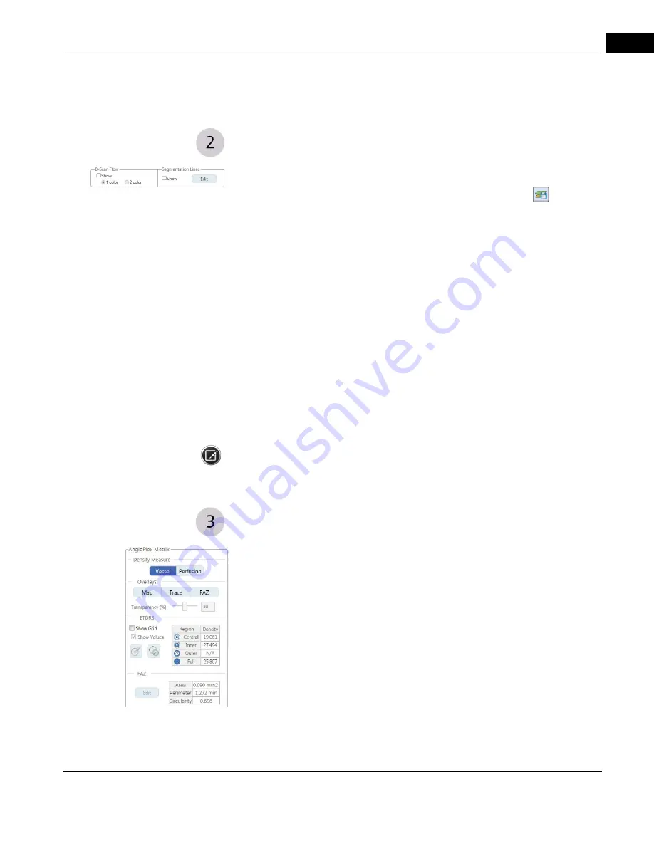
• B-scan Flow (one color or two color): Selecting this option adds one or two colors to
the B-scan viewport. Selecting
One Color
will show all aspects of the flow in light red.
Choosing
Two Color
will overlay the scan such that the light red will overlay the data
lying above the RPE and green will overlay data lying below the RPE.
• Segmentation Lines: Selecting this option adds the dashed magenta lines to the
segment viewport (lower left).
1.Select Edit.
2.Left-click the mouse at an end (where the triangle is) of one of the lines and
hold down the mouse button to move it. This will change the offset and define a
new slab.
These changes will be reflected in the AngioPlex and Structure images above. You
cannot use segmentation lines when the images are overlaid with a thickness map.
NOTE: This option is not available for Montage Angio.
AngioPlex Metrix
AngioPlex Metrix provides quantification for various OCTA parameters. The appearance of
the AngioPlex Metrix for Angiography looks like the example margin graphic on the left.
The appearance of the AngioPlex Metrix for ONH Angiography looks like the example
margin graphic on the following page. The AngioPlex Metrix is available for the
following scans:
• Angiography 6 mm x 6 mm
• Angiography 3 mm x 3 mm
• ONH Angiography 4.5 mm x 4.5 mm
Summary of Contents for CIRRUS HD-OCT 500
Page 1: ...2660021156446 B2660021156446 B CIRRUS HD OCT User Manual Models 500 5000 ...
Page 32: ...User Documentation 2660021169012 Rev A 2017 12 CIRRUS HD OCT User Manual 2 6 ...
Page 44: ...Software 2660021169012 Rev A 2017 12 CIRRUS HD OCT User Manual 3 12 ...
Page 58: ...User Login Logout 2660021169012 Rev A 2017 12 CIRRUS HD OCT User Manual 4 14 ...
Page 72: ...Patient Preparation 2660021169012 Rev A 2017 12 CIRRUS HD OCT User Manual 5 14 ...
Page 110: ...Tracking and Repeat Scans 2660021169012 Rev A 2017 12 CIRRUS HD OCT User Manual 6 38 ...
Page 122: ...Criteria for Image Acceptance 2660021169012 Rev A 2017 12 CIRRUS HD OCT User Manual 7 12 ...
Page 222: ...Overview 2660021169012 Rev A 2017 12 CIRRUS HD OCT User Manual 9 28 ...
Page 256: ...Log Files 2660021169012 Rev A 2017 12 CIRRUS HD OCT User Manual 11 18 ...
Page 308: ...Appendix 2660021169012 Rev A 2017 12 CIRRUS HD OCT User Manual A 34 ...
Page 350: ...CIRRUS HD OCT User Manual 2660021169012 Rev A 2017 12 I 8 ...
Page 351: ...CIRRUS HD OCT User Manual 2660021169012 Rev A 2017 12 ...