GE
V
OLUSON
i / V
OLUSON
e
D
IRECTION
KTI106052, R
EVISION
10
S
ERVICE
M
ANUAL
5-16
Section 5-2 - General Information
5-2-5-9
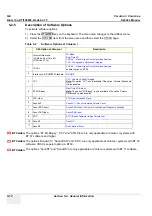
SonoAVC - Sono Automated Volume Count
This Feature can automatically detect low echogenic objects (e.g., follicles) in a volume of an organ
(e.g., ovary) and analyze their shape and volume. From the calculated volume an average diameter can
be calculated. It also lists the objects according to their size.
“Separation” is controlling a parameter of the segmentation algorithm that defines an initial threshold to
separate objects. Increasing this parameter will prevent objects from being identified as multiple objects
(e.g. when there is noise within the object), but might prevent small objects from being found correctly.
“Growth” is controlling a parameter in the segmentation algorithm that defines the final shape of the
objects found. Increasing this parameter will allow the objects to fit tighter to the visible boundary. A
value too large might cause the objects to grow over the boundary and cover areas no longer part of
the objects of interest.
5-2-5-10
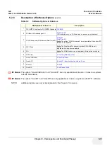
SonoVCAD Heart- Computer Assisted Heart Diagnosis Package
VCAD is a technology that automatically generates a number of views of the fetal heart to make
diagnosis easier. At this time it can help to find the right and left outflow tract of the heart and the fetal
stomach.
5-2-5-11
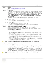
SonoVCAD labor
VCAD labor allows for supervision of labor using specific measurements aided by onscreen orientation
marks.
This software has the ability to electronically edit the images, and makes it possible to cut away
“3D artifacts”.
5-2-5-12
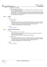
STIC (Spatio-Temporal Image Correlation)
With this acquisition method the fetal heart or an artery can be visualized in 4D.
It is not a Real Time 4D technique, but a post processed 3D acquisition.
In order to archive a good result, try to adjust the size of the volume box and the sweep angle to be as
small as possible. The longer the acquisition time, the better the spatial resolution will be.
A good STIC, STIC CFM (2D+CFM), STIC PD (2D+PD) or STIC HD (2D+HD-Flow) data set shows a
regular and synchronous pumping of the fetal heart or of an artery.
The user must be sure that there is minimal movement of the participating persons (e.g., mother and
fetus), and that the probe is held absolutely still throughout the acquisition period. Movement will cause
a failure of the acquisition. The acquired images are post processed to calculate a 4D Volume Cine
sequence. Please make sure that the borders of the fetal heart or the artery are smooth and there are
no sudden discontinuities. If the user (trained operator) clearly recognizes a disturbance during the
acquisition period, the acquisition has to be cancelled.
• STIC - Fetal Cardio is only available on RAB & RIC probes in the OB/GYN application.
• STIC - Vascular is only available on the RSP probe in the Peripheral Vascular application.
BT
Version:
BT-Version:
The option “SonoAVC” is only applicable at Voluson i / Voluson e systems with´BT´09 software.
BT
Version:
BT-Version:
The option “SonoVCAD Heart” is only applicable at Voluson i with BT´09 software.
BT
Version:
BT-Version:
The option “SonoVCAD labor” is only applicable at Voluson i / Voluson e systems with BT´09 software.
BT
Version:
BT-Version:
The option “STIC” is only applicable at Voluson i systems with´BT´09 software.
Summary of Contents for Voluson i BT06
Page 2: ......
Page 11: ...GE VOLUSON i VOLUSON e DIRECTION KTI106052 REVISION 10 SERVICE MANUAL ix ZH CN KO...
Page 44: ...GE VOLUSON i VOLUSON e DIRECTION KTI106052 REVISION 10 SERVICE MANUAL xlii Table of Contents...
Page 514: ...GE VOLUSON i VOLUSON e DIRECTION KTI106052 REVISION 10 SERVICE MANUAL IV Index...
Page 515: ......

