


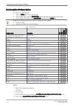


5.1.1 Description of Operating Modes
5.1.1.1 B-Mode or 2D-Mode
B-Mode or 2D-mode is a two-dimensional image of the amplitude of the echo signal. It is used for location
and measurement of anatomical structures and for spatial orientation during operation of other modes. In
2D-mode, a two-dimensional cross-section of a three-dimensional soft tissue structure such as the heart is
displayed in real time. Ultrasound echoes of different intensities are mapped to different gray scale or color
values in the display. The outline of the 2D cross-section may be a rectangle, parallelogram, sector or 360-
degree circle, depending on the transducer used. 2D-mode can be used in combination with any other mode.
5.1.1.1.1 Coded Harmonic Imaging (HI)
In Harmonic Imaging, acoustic aberrations due to tissue are minimized by receiving and processing the
second harmonic signal that is generated within the insonified tissue. Voluson E-Series high performance HI
provides superb detail resolution and penetration, outstanding contrast resolution, excellent acoustic clutter
rejection and an easy to operate user interface. Coded Harmonics enhances near field resolution for
improved small parts imaging as well as far field penetration. It diminishes low frequency amplitude noise
and improves imaging technically difficult patients. It may be especially beneficial when imaging isoechoic
lesions in shallow-depth anatomy in the breast, liver and hard-to-visualize fetal anatomy. Coded Harmonics
may improve the B-Mode image quality without introducing a contrast agent.
5.1.1.1.2 XTD-View
XTD-View (Extended View) provides the ability to construct and view a static 2D image which is wider than
the field of view of a given probe. This feature allows viewing and measurement of anatomy that is larger
than what would fit in a single image. XTD-View constructs the extended image from individual image frames
as the operator slides the probe along the surface of the skin in direction of the scan plane. Examples
include scanning of vascular structures and connective tissues in the arms and legs.
5.1.1.1.3 B-Flow
Section 5.1.4.6 "B-Flow" on page 5-14
5.1.1.1.4 Coded Contrast Imaging (optional)
Section 5.1.4.7 "Coded Contrast Imaging" on page 5-14
.
5.1.1.1.5 Elastography (optional)
Section 5.1.4.16 "Elastography" on page 5-17
.
5.1.1.2 M-Mode
In M-mode, soft tissue structure is presented as scrolling display, with depth on the Y-axis and time on the X-
axis. It is used primarily for cardiac measurements such as value timing on septal wall thickness when
accurate timing information is required. M-mode is also known as T-M mode or time-motion mode.
Ultrasound echoes of different intensities are mapped to different gray scale values in the display. M-mode
displays time motion information of the ultrasound data derived from a stationary beam. Depth is arranged
along the vertical axis with time along the horizontal axis. M-mode is normally used in conjunction with a 2D
image for spatial reference. The 2D image has a graphical line (M-line) superimposed on the 2D image
indicating where the M-mode beam is located.
5.1.1.2.1 MCFM Mode (M Mode + Color Flow Mode)
Color Flow Mode and Color M Mode are Doppler modes intended to add color-coded qualitative information
concerning the relative velocity and direction of fluid motion within the 2D mode or M mode image. Color
Flow overlays color on the M mode trace using velocity and variance color maps. The Color Flow wedge
overlays the 2D mode image and M mode timeline.
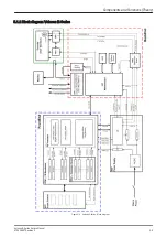
Components and Functions (Theory)
5-6
Voluson E-Series Service Manual
KTD106657 Revision 2
Summary of Contents for H48681XB
Page 11: ...Introduction Voluson E Series Service Manual KTD106657 Revision 2 1 3 ...
Page 12: ...Introduction 1 4 Voluson E Series Service Manual KTD106657 Revision 2 ...
Page 13: ...Introduction Voluson E Series Service Manual KTD106657 Revision 2 1 5 ...
Page 14: ...Introduction 1 6 Voluson E Series Service Manual KTD106657 Revision 2 ...
Page 15: ...Introduction Voluson E Series Service Manual KTD106657 Revision 2 1 7 ...
Page 16: ...Introduction 1 8 Voluson E Series Service Manual KTD106657 Revision 2 ...
Page 17: ...Introduction Voluson E Series Service Manual KTD106657 Revision 2 1 9 ...
Page 365: ......
Page 366: ...GE Healthcare Austria GmbH Co OG Tiefenbach 15 4871 Zipf Austria www gehealthcare com ...