


5.1.4.15 SonoBiometry
SonoBiometry is an alternative to the common fetal biometry measurements. It provides system suggested
measurements for BPD, HC, AC and FL which need to be confirmed by the user or can be changed
manually.
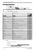
The following automated fetal biometry measurements are available:
•
BPD (o-o) – Biparietal diameter type: outside-outside
•
BPD (o-i) - Biparietal diameter type: outside-inner
•
HC – Head circumference
•
AC – Abdomen circumference
•
FL – Femur length
•
BPD + HC: combined measurement
The measurement mode can be changed from automatic to manual. Available measurement methods
depend on this selection and on the measurement item itself.
It is not necessary to select a region where the measurement should be performed
5.1.4.16 Elastography
Elastography refers to the measurement of elastic properties of tissues, based on the well-established
principle that malignant tissue is harder than benign tissue.
Elastography shows the spatial distribution of tissue elasticity properties in a region of interest by estimating
the strain before and after tissue distortion caused by external or internal forces. The strain estimation is
filtered and scaled to provide a smooth presentation when displayed.
During scanning in the elastography mode, the examiner manually slightly compresses the tissue using the
ultrasound probe. A strain correlation (strain is the deformation of the tissue by compression) is continuously
performed for visual perception on the monitor.
5.1.4.16.1 Elastography Analysis
A selectable sequence of Elastography images are analyzed within a ROI (range of interest). The Strain %
or the Elasticity Index is displayed as curves over the time.
The mean value of the Strain % is measured within 1 or more ROI’s and Ratios are calculated. “Generic
Elasto” measurements are located in the generic measurement menu and are only available if
“Elastography” is activated.
5.1.4.17 SRI Advanced - Speckle Reduction Imaging
A type of image noise or interference is generally considered undesirable and can obscure the quality or
interpretation of B-mode images. Although somewhat associated with the underlying echogenicity of tissue
scatters, image speckle characteristics such as brightness, density or size have no apparent value in
determining tissue structure or related properties. The elimination of or significant reduction in speckle
improves the quality or diagnostic potential of the image. The method applied in the subject modification
utilizes a nonlinear diffusion filtering technique that permits effective speckle reduction in real time. The
speckle reduction filter is available to the user in all B-mode imaging, independent of the used probe.
5.1.4.18 V-SRI Advanced - Speckle Reduction Imaging
V-SRI is an enhancement of the existing SRI algorithms and improves specially the C-plane image. This new
algorithm (from Context Vision) is used in the A-, B-, C-planes and rendered images instead of the
conventional SRI.
Note
The “V-SRI” option is only available at Voluson E8 Expert systems with probe RM6C, RIC5-9-D and
RIC6-12-D.
5.1.4.19 CW - Continuous Wave Doppler
Section 5.1.5.1 "CW - Continuous Wave Doppler" on page 5-18
Note
Additional hardware required!
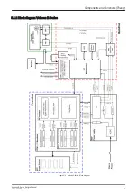
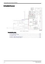
Components and Functions (Theory)
Voluson E-Series Service Manual
KTD106657 Revision 2
5-17
Summary of Contents for H48681XB
Page 11: ...Introduction Voluson E Series Service Manual KTD106657 Revision 2 1 3 ...
Page 12: ...Introduction 1 4 Voluson E Series Service Manual KTD106657 Revision 2 ...
Page 13: ...Introduction Voluson E Series Service Manual KTD106657 Revision 2 1 5 ...
Page 14: ...Introduction 1 6 Voluson E Series Service Manual KTD106657 Revision 2 ...
Page 15: ...Introduction Voluson E Series Service Manual KTD106657 Revision 2 1 7 ...
Page 16: ...Introduction 1 8 Voluson E Series Service Manual KTD106657 Revision 2 ...
Page 17: ...Introduction Voluson E Series Service Manual KTD106657 Revision 2 1 9 ...
Page 365: ......
Page 366: ...GE Healthcare Austria GmbH Co OG Tiefenbach 15 4871 Zipf Austria www gehealthcare com ...