68
3.
Reconstitute one vial from the Low pI, High pI or Broad pI
calibration kit in 30-40 µl of distilled water for Coomassie stain-
ing, and in 2 ml for silver staining. Reconstituted kit proteins can
be store frozen at -20°C.
4.
Start the run and apply the samples to the gel (use the
procedures presented in Separation procedures, section 5.2).
Apply calibration proteins between the sample lanes. On one
side of the calibration proteins, apply the sample at the cathode,
and on the other side at the anode. This will help to ensure that
proteins are in equilibrium for pI measurements (they should
focus at the same point in the gradient).
5.
After electrophoresis, develop the protein bands according to one
of the development methods given in chapter 9,
Develop-
ment Technique Files.
6.
To easily measure the band distance, mount the gel in a slide
frame and project the image to the desired format using a slide
projector. Alternatively, scan the gel.
7.
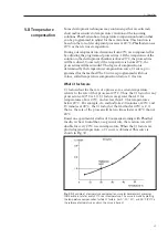
Plot the known pI value of each pI calibration protein versus its
distance from a reference point, e.g. the cathode (to the nearest
0.05 cm). Draw a line through the points to obtain the pH
gradient profile of the gel.
8.
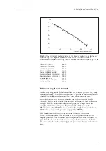
Measure the distance from the reference to the proteins of
interest. Use the pH profile of the gel to interpolate the pI
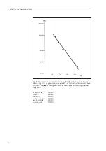
points of these proteins. Figure 1 shows an example of a pH
gradient profile established using the Broad pI calibration kit.
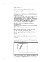
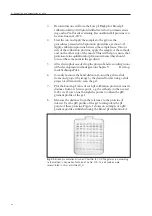
Fig. 33.
Broad pI calibration kit run on PhastGel IEF 3-9 The gel was run according
to the method in Separation Technique File No. 100. The kit proteins were
reconstituted in 35 µl of distilled H
2
O.
6. Evaluation and presentation of data
Содержание PhastSystem
Страница 1: ...Phast System user manual automated electrophoresis um 80 1320 15 Edition AI ...
Страница 2: ......
Страница 8: ...8 ...
Страница 30: ...30 ...
Страница 34: ...34 ...
Страница 64: ...64 ...
Страница 96: ...12 ...
Страница 104: ......
Страница 105: ......
Страница 106: ...PRINTED IN SWEDEN BY TK I UPPSALA AB 2003 ...

