GE
V
OLUSON
i / V
OLUSON
e
D
IRECTION
KTI106052, R
EVISION
10
S
ERVICE
M
ANUAL
4-16
Section 4-4 - Functional Checks
4-4-5-1
Pre-Volume Mode Functions
(cont’d)
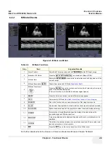
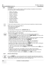
Table 4-7
Pre-Volume Mode Functions
Step
Task
Expected Results
1
3D RENDERING
3D Static volume acqui rendered 3D image (also in combination Color or PD Mode)
2
SECTIONAL PLANES
3D Static volume acquisition resp. 4D volume acquisition without rendered 3D image
3
TUI
*
This method of visualization is consistent with the way other medical systems such as CT or
MRI, present the data to the user (slices through the data set, which are parallel to each other).
4
4D REAL TIME
Real Time 4D - continuous volume acquisition and parallel calculation of 3D rendered images
5
RT 4D BIOPSY*
BT
Version:
BT-Version:
Real Time 4D Biopsy
continuous volume acquisition and parallel calculation of 3D rendered images
This feature is only available at Voluson i systems with
BT´07
software and higher.
6
VCI*
BT
Version:
BT-Version:
Volume Contrast Imaging - improves the contrast resolution and the signal / noise ratio and
therefore facilitates finding of diffuse lesions in organs
This feature is only available at Voluson i systems with
BT´07
software and higher.
7
STIC*
BT
Version:
BT-Version:
The fetal heart or an artery can visualized in 4D (also in combination with PD, HD-Flow or CFM)
This feature is only available at Voluson i systems with BT´09 software.
8
SONOVCAD HEART*
BT
Version:
BT-Version:
VCAD is a technology that automatically generates a number of views of the fetal heart to make
diagnosis easier. At this time it can help to find the right and left outflow tract of the heart and
the fetal stomach. By aligning the 4 Chamber view of the heart to a set diagram the system will
generate views of LVOT, RVOT, Stomach, IVC/SVC and Ductal arch
This feature is only available at Voluson i systems with BT´09 software.
9
SONOVCAD LABOR
BT
Version:
BT-Version:
This Feature allows for supervision of labor using specific measurements aided by onscreen
orientation marks.
This feature is only available at Voluson i / Voluson e systems with BT´09 software.
10
SONOAVC
BT
Version:
BT-Version:
This Feature can automatically detect low echogenic objects (e.g., follicles) in a volume of an
organ (e.g., ovary) and analyze their shape and volume
This feature is only available at Voluson i / Voluson e systems with BT´09 software.
11
CLASSIC: Advanced Mode with dedicated Voluson Volume features
SMART: Easy/Speedy operation with as few controls as possible
12
- Quarter size display of Sectional Planes without 3D image
or
- Quarter size display of Sectional rendered 3D image
(Note: The display depends on selected Acquisition- and Visualization Mode!)
13
Dual size display of Sectional rendered 3D image.
(Note: The display depends on selected Acquisition- and Visualization Mode!
This format is not possible for Static 3D Acquisition)
14
- Full size display of a the reference image
or
- Full size display of the rendered 3D image.
(Note: The display depends on selected Acquisition- and Visualization Mode!)
15
Volume Box Position
and Volume Box Size
Adjust the POS (Position) resp. SIZE of the Volume Box (ROI = Region Of Interest) with the
trackball in the 2D Single image.
The
upper trackball key
to change the trackball function from Box Position to Box Size.
16
QUALITY
Changes the line density against the acquisition speed (low, mid1, mid2, high1, high2).
17
VOL. ANGLE
To select the Volume Sweep Angle.
18
Start Acquisition
Press the
FREEZE
key resp. the
right trackball key
to start the Volume acquisition.
Summary of Contents for H48651KR
Page 2: ......
Page 11: ...GE VOLUSON i VOLUSON e DIRECTION KTI106052 REVISION 10 SERVICE MANUAL ix ZH CN KO ...
Page 44: ...GE VOLUSON i VOLUSON e DIRECTION KTI106052 REVISION 10 SERVICE MANUAL xlii Table of Contents ...
Page 514: ...GE VOLUSON i VOLUSON e DIRECTION KTI106052 REVISION 10 SERVICE MANUAL IV Index ...
Page 515: ......

