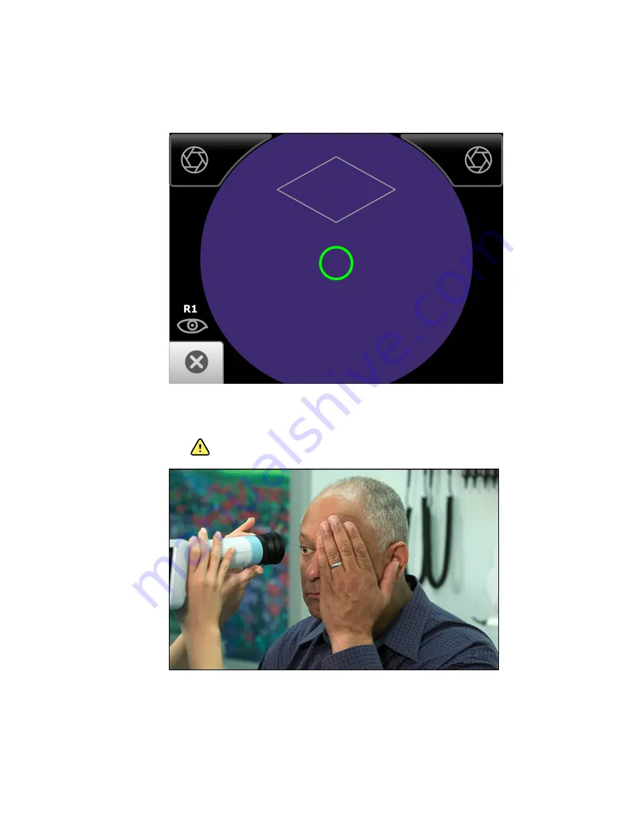
4. Touch
Start
to enter the
Image acquisition mode
and begin the first exam of the
patient's right eye (R1).
The
Exam acquisition screen
appears.
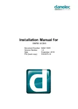
5. Hold the patient end of the RetinaVue 100 Imager two to three inches directly in
front of the patient's examined eye. Continue forward to compress the eye cup
against the examined eye.
WARNING
Clean and disinfect the eye cup after each
patient to avoid the risk of cross-contamination.
6. Direct the patient to focus on the green fixation lights inside the barrel of the
RetinaVue 100 Imager.
Note
Instruct the patient to cover, but not close, their unexamined
eye. This will help the patient to focus on the green fixation
lights.
50 Using the RetinaVue 100 Imager
Welch Allyn RetinaVue™ 100 Imager
Содержание RetinaVue 100 Imager
Страница 1: ...Welch Allyn RetinaVue 100 Imager Directions for use Software version 6 XX...
Страница 8: ...4 Symbols Welch Allyn RetinaVue 100 Imager...
Страница 14: ...10 Introduction Welch Allyn RetinaVue 100 Imager...
Страница 59: ...Directions for use Using the RetinaVue 100 Imager 55...
Страница 86: ...82 General compliance and standards Welch Allyn RetinaVue 100 Imager...
Страница 112: ...108 Appendix Welch Allyn RetinaVue 100 Imager...
Страница 114: ......
Страница 115: ......
Страница 116: ...Material No 411492...


