Acquire Screen and Controls
2660021169012 Rev. A 2017-12
CIRRUS HD-OCT User Manual
6-24
Fundus Viewport
The Fundus Viewport lies in the lower left quadrant of the main Acquire screen Viewport as
shown in
, and shows a live fundus image from the line scanning
ophthalmoscope (LSO). The Fundus Viewport is not available for Anterior scans. As with
the Iris Viewport, there are a number of options available for optimizing the patient scan:
• Focus/Auto Focus will attempt to compensate for the patient’s refractive error by
automatically changing the focus adjustment. This may help clear up a dim fundus
view and will also help clear up the fixation target for a patient whose refractive error
is considerable. In addition to improving the overall focus, the Auto Focus feature will
do an additional adjustment on the brightness and contrast of the fundus image.
• The Transparency slider is active when a saved scan image overlay is present, which
occurs when you are using a prior scan.
• Prior scan will appear as a selectable link, as shown in the margin. If your patient has
been scanned previously, selecting this option will open up a small screen with a list of
the patient’s previous scans. Once you select a previous scan, the location of that scan
will appear as a live link, and serve as a reference as you acquire the current scan. You
can change the prior, reference scan, at any time. You can also initiate an exact replica
of a prior scan using the Auto Repeat function (
• Optimize automatically optimizes first the scan image centering (Z-offset), and then
optimizes the scan image quality (polarization). Instruct the patient not to blink during
optimization.
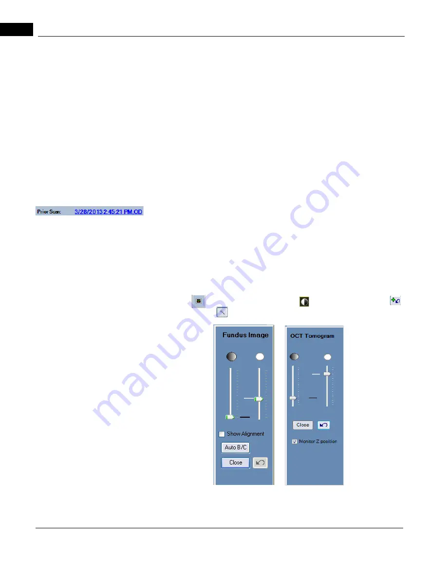
• Enhance (
), adjust brightness and contrast (
), or reset the fixation target
.
When you select (
), one of the following dialog boxes will appear.
• If you have the tracking selection enabled (
tracking on the macula, FastTrac automatically monitors whether the OCT B-scans are
Содержание CIRRUS HD-OCT 500
Страница 1: ...2660021156446 B2660021156446 B CIRRUS HD OCT User Manual Models 500 5000 ...
Страница 32: ...User Documentation 2660021169012 Rev A 2017 12 CIRRUS HD OCT User Manual 2 6 ...
Страница 44: ...Software 2660021169012 Rev A 2017 12 CIRRUS HD OCT User Manual 3 12 ...
Страница 58: ...User Login Logout 2660021169012 Rev A 2017 12 CIRRUS HD OCT User Manual 4 14 ...
Страница 72: ...Patient Preparation 2660021169012 Rev A 2017 12 CIRRUS HD OCT User Manual 5 14 ...
Страница 110: ...Tracking and Repeat Scans 2660021169012 Rev A 2017 12 CIRRUS HD OCT User Manual 6 38 ...
Страница 122: ...Criteria for Image Acceptance 2660021169012 Rev A 2017 12 CIRRUS HD OCT User Manual 7 12 ...
Страница 222: ...Overview 2660021169012 Rev A 2017 12 CIRRUS HD OCT User Manual 9 28 ...
Страница 256: ...Log Files 2660021169012 Rev A 2017 12 CIRRUS HD OCT User Manual 11 18 ...
Страница 272: ...Electrical Physical and Environmental 2660021169012 Rev A 2017 12 CIRRUS HD OCT User Manual 13 4 ...
Страница 292: ...Appendix 2660021169012 Rev A 2017 12 CIRRUS HD OCT User Manual A 18 cáÖìêÉ JV kçêã íáîÉ a í aÉí áäë oÉéçêí ...
Страница 308: ...Appendix 2660021169012 Rev A 2017 12 CIRRUS HD OCT User Manual A 34 ...
Страница 350: ...CIRRUS HD OCT User Manual 2660021169012 Rev A 2017 12 I 8 ...
Страница 351: ...CIRRUS HD OCT User Manual 2660021169012 Rev A 2017 12 ...


