Chapter A5: Image Viewer
Definium AMX 700 X-Ray System
5161515-1EN
Rev. 6 (10 February 2008)
A5-11
Operator Manual
© 2008 General Electric Company. All rights reserved.
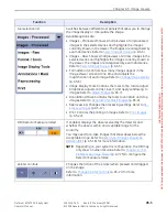
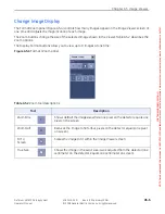
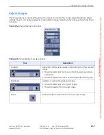
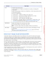
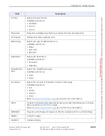
Full
Places all available system annotation on the image.
• Patient information (top left corner) exam date and patient
identification
• Study information (top left corner) exam identification
• Series information (top left corner) series identification
• Image information (top left corner) image identification
• Acquisition information (top right corner) dose
• Hospital information (top right corner) the name of the facility where
the image was acquired
• X-ray parameters (top right corner) the mA, kVp, ms, mAs, and DEI of
the exposure
• Anatomy information (bottom left corner) the protocol used to acquire
the image
• Processing information (bottom left corner) the look used to process
the image
• User measurements (bottom right corner) size and angle
measurements for line, ellipse, and Cobb annotations
Display parameters (bottom right corner) the size of the image and the
zoom
Partial
Displays ONLY the facility name, dose information and technical factors.
None
Removes all system annotations from the image. System annotations can
be re-applied by pressing [FULL], [PARTIAL], or [CUSTOM].
Custom
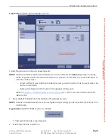
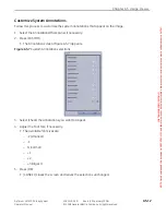
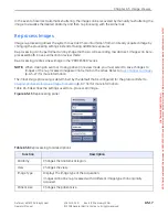
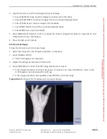
Brings up a screen (Figure A5-7) that allows you to choose which system
annotations appear. Refer to
Manual
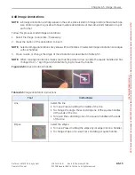
Shutter
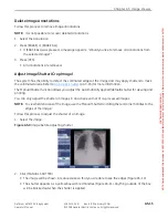
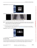
Manually adjusts the image shutter.
Collimation is detected using image-based processing. In some cases, the
FOV detected by the system does not match the actual exposed FOV. Use
the Manual Shutter tool to correct this.
NOTE:
This function is only available when the image is open in a live exam
or for re-processed images.
Refer to
Adjust Image Shutter (Crop Image)
(p. A5-15) for more information.
Tool
Description
FOR
TRAINING
PURPOSES
ONLY!
NOTE:
Once
downloaded,
this
document
is
UNCONTROLLED,
and
therefore
may
not
be
the
latest
revision.
Always
confirm
revision
status
against
a
validated
source
(ie
CDL).

