CHAP
. 2
GE Healthcare
Senographe 2000 D Acquisition System
REV 1
OM 5179217–1–100
23
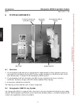
Images are acquired by direct digitization; they are displayed immediately on the AWS monitor and are
stored for later diagnostic review. They can be processed and/or filmed.
AOP (Automatic Optimization of Parameters) and manual setting modes are provided for control of
X-ray parameters; the system provides auto-collimation and other leading features.
8-3
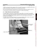
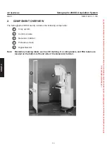
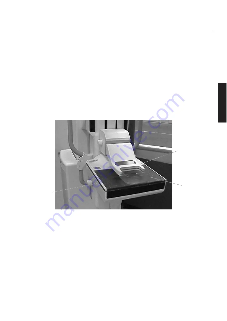
Digital Detector and Image Receptor
The Digital Detector is built into the Image Receptor, shown below. It is a flat panel of amorphous
silicon on which cesium iodide is deposited to maximize detection of X-Rays and transmission of light
photons. The high definition digital images produced are sent to the Acquisition Workstation for
visualization and processing.
The upper surface of the Image Receptor is a removable grid (Bucky); when the grid is not required, it
is easily removed and replaced by an optional breast holder without grid.
Image Receptor
Compression
Paddle
Bucky
FOR
TRAINING
PURPOSES
ONLY!
NOTE:
Once
downloaded,
this
document
is
UNCONTROLLED,
and
therefore
may
not
be
the
latest
revision.
Always
confirm
revision
status
against
a
validated
source
(ie
CDL).


