CHAP
. 10
GE Healthcare
Senographe 2000 D Acquisition System
REV 1
OM 5179217–1–100
114
4-1
Use of Markers in AOP Mode
The algorithm used in AOP mode searches for the most dense part of the breast, and uses this as a
reference in its calculations. It is therefore important to avoid the presence of dense objects in the
area used by the algorithm.
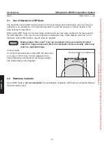
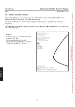
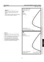
When using AOP mode, do not place large markers such as view name markers in the area used by
the AOP algorithm. They may be used anywhere outside this area. Small markers less than 3 mm
diameter, such as BB markers, may be used as required.
Markers larger than 2 mm
2
must not be present in the area used by the AOP
algorithm. Large markers will affect the calculation of tissue density, which may
lead to a degraded image.
Contact mode:
In contact mode exposures using AOP, the area used
is an area of 160 mm by 140 mm adjacent to the
chest wall side and centered on the image receptor
(the shaded area in the diagram).
4-2
Mammary implants
Use of AOP mode is
not
recommended
for examinations of patients with mammary implants. Manual
mode should be used.
CAUTION
AOP ROI
140 mm
160 mm
No large markers in shaded area
FOR
TRAINING
PURPOSES
ONLY!
NOTE:
Once
downloaded,
this
document
is
UNCONTROLLED,
and
therefore
may
not
be
the
latest
revision.
Always
confirm
revision
status
against
a
validated
source
(ie
CDL).

