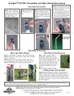
Safety measures
OPMI Lumera i
Version 8.0
Page 16
G-30-1720-en
Measures to prevent phototoxic injury
Several papers
1)-5)
dealing with the problem of phototoxicity in ophthalmic
surgery have been published. A comprehensive review of these publications
reveals five aspects of particular concern:
–
Illumination characteristics (spectral composition)
–
Illumination intensity
–
Angle of illumination
–
Focus of the light source
–
Exposure time to light
In the following, comments on these aspects are given and a description of
how ZEISS, as a manufacturer, makes allowance for them in its systems.
Illumination characteristics (spectral composition)
Studies on exposing the eye to light with varying spectral compositions have
been conducted since as early as the 1950s. These studies suggest that the
potential hazard of phototoxic injury to the patient’s retina can be reduced by
blocking out the blue and ultraviolet light below a wavelength of 475 nm.
Integrated protection filters
For protection of the retina, ZEISS offers the swing-in retina protection filter
(blue barrier filter) and the stationary UV blocking filter as standard features
of the surgical microscope. This reduces not only the exposure of the patient’s
eye to light, but also that of the surgeon.
An important point to note here, however, is that the use of filters will
inevitably change the perceived color of the light. For this reason, the surgeon
may initially have to get used to the changed appearance of the anatomical
structures.
Содержание OPMI Lumera i
Страница 1: ...ZEISS OPMI Lumera i on the floor stand Instructions for Use G 30 1720 en Version 8 0 2018 11 26...
Страница 4: ...OPMI Lumera i Version 8 0 Page 4 G 30 1720 en...
Страница 25: ...Version 8 0 G 30 1720 en Page 25 OPMI Lumera i Safety measures Fig 2 Switch for manual mode 3 1 3...
Страница 32: ...Safety measures OPMI Lumera i Version 8 0 Page 32 G 30 1720 en...
Страница 35: ...Version 8 0 G 30 1720 en Page 35 OPMI Lumera i Device overview Fig 4 System overview 3 1 2...
Страница 37: ...Version 8 0 G 30 1720 en Page 37 OPMI Lumera i Device overview Fig 5 Components of the microscope 2 2 1 4 3...
Страница 39: ...Version 8 0 G 30 1720 en Page 39 OPMI Lumera i Device overview Fig 6 Controls on the microscope 2 3 4 1...
Страница 51: ...Version 8 0 G 30 1720 en Page 51 OPMI Lumera i Device overview Fig 15 Connectors on the floor stand 9 10 11 12...
Страница 61: ...Version 8 0 G 30 1720 en Page 61 OPMI Lumera i Preparation for use...
Страница 71: ...Version 8 0 G 30 1720 en Page 71 OPMI Lumera i Preparation for use Fig 24 C mount adapter 3 7 4 2 1 5 8 6...
Страница 83: ...Version 8 0 G 30 1720 en Page 83 OPMI Lumera i Preparation for use...
Страница 88: ...Preparation for use OPMI Lumera i Version 8 0 Page 88 G 30 1720 en...
Страница 97: ...Version 8 0 G 30 1720 en Page 97 OPMI Lumera i Operation...
Страница 99: ...Version 8 0 G 30 1720 en Page 99 OPMI Lumera i Operation Fig 35 Menu structure 2 3 7 6 5 4 1 8...
Страница 149: ...Version 8 0 G 30 1720 en Page 149 OPMI Lumera i Device data Fig 39 Dimensional drawing of OPMI Lumera i...
Страница 182: ...OPMI Lumera i Version 8 0 Page 182 G 30 1720 en...
Страница 183: ...Version 8 0 G 30 1720 en Page 183 OPMI Lumera i Blank page for your notes...