


DM DOC 040 Rev 1.0
Resting ECG general description
Electrocardiography (ECG or EKG from Greek: kardia, meaning heart) is a noninvasive
transthoracic interpretation of the electrical activity of the heart over a period of time. D-MAS
detects and records ECG signals with electrodes attached to the surface of the skin. The
recording produced by this procedure is termed an electrocardiogram (also ECG or EKG).
The ECG is the most important test for interpretation of the cardiac rhythm, conduction system
abnormalities, and for the detection of myocardial ischemia. The ECG is also of great value in the
evaluation of other types of cardiac abnormalities including valvular heart disease,
cardiomyopathy, pericarditis, and hypertensive disease. Finally, the ECG can be used to monitor
drug treatment (specifically antiarrhythmic therapy) and to detect metabolic disturbances
Principle of operation
The basic principle of the ECG is that stimulation of a muscle alters the electrical potential of the
muscle fibres. Cardiac cells, unlike other cells, have a property known as automaticity, which is
the capacity to spontaneously initiate impulses. These are then transmitted from cell to cell by
gap junctions that connect cardiac cells to each other.
Electrical impulses spread through the muscle cells because of changes in ions between
intracellular and extracellular fluid. This is referred to as action potential. The primary ions
involved are potassium, sodium and calcium. The action potential is the potential for action
created by the balance between electrical charges (positive and negative) of ions on either side of
the cell membrane.
When the cells are in a resting state, the insides are negatively charged compared to the
outsides. Membrane pumps act to maintain this electrical polarity (negative charge) of the cardiac
cells. Contraction of the heart muscle is triggered by depolarisation, which causes the internal
negative charge to be lost transiently.
However, following depolarisation, the cardiac cells return again to their resting charge, known as
repolarisation.
These waves of depolarisation and repolarisation represent an electrical current and can be
detected by placing electrodes on the surface of the body. After the current has spread from the
heart through the body, the changes are picked up by electrodes connected to D-MAS and the
activity is recorded digitally. The D-MAS ECG is therefore a graphic representation of the
electrical activity in the heart. The current is transmitted at the pre-defined points of contact of the
electrode with the body.
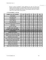
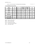
The ECG comprises 12 leads: three standard leads (lead I, lead II and lead III); three augmented
leads (AVR, AVL and AVF); and six chest leads (V1, V2, V3, V4, V5, V6).
A total of 10 electrodes (points of contact with the body) are used to perform the ECG.
It is called a 12 lead ECG because the heart is viewed and recorded from 12 different viewpoints.
Two electrodes are placed on the arms and two on the legs to provide the basis for the six limb
leads (three standard and three augmented leads).
The four limb electrodes record six different views of electrical activity in the heart, while each of
the chest electrodes records one lead each.
The ECG signals are acquired via electrodes attached to the ECG patient cable. The signals are
digitized, displayed and stored electronically. The recorded signal is measured and an ECG
interpretation algorithm is applied to the recording providing the user with displayed
measurements and interpretation statements.
© 2013 DanMedical Ltd
23


