GE H
EALTHCARE
DRAFT
V
OLUSON
E8 / V
OLUSON
E6
D
IRECTION
KTD102576, R
EVISION
7
DRAFT (A
UGUST
23, 2012)
S
ERVICE
M
ANUAL
Chapter 5 - Components and Functions (Theory)
5-19
5-2-4-11
Anatomical M-Mode (AMM)
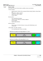
Anatomical M-Mode displays a distance/time plot from a cursor line, which can be defined freely.
The M-Mode display changes according to the motion of the M cursor. In the Dual format, two defined
distances can be displayed at the same time.
AMM is available in grayscale and color modes (CF, HD Flow, TD)
•
simultanous Display of 2 M-Mode Cursors in 2D Mode
•
each Cursor is freely rotatable
•
can be done after Freeze and on reloaded Cine
5-2-4-12
Scan Assistant
All major ultrasound societies (SMFM, AIUM, ACR, ACOG) have guidelines to be followed for each
examination. For legal reasons, documentation is recommended for all structures evaluated.
Scan Assistant prevents the user from missing an important step of an examination. Completely
customizable, each exam can have sub menus that allow measurements and annotations to be tagged
for transfer with DICOM and DICOM SR.
5-2-4-13
Advanced STIC (Spatio-Temporal Image Correlation)
5-2-4-13-1
Basic STIC
5-2-4-13-2
STIC M-Mode
Creates a M-spectrum from a STIC acquisition (capture of a full fetal heart cycle in real-time saved
as a volume for later analysis). After activating STIC M-Mode the STIC cine is running and the STIC
M-spectrum will be shown. In STIC-M all M-Measurements are possible.
Furthermore the M-cursor is available as a freeform line type.
-
can be done in A, B or C Plane and can be done postprocessing
-
possibility to perform measurements for evaluation of ventricle contraction
-
possibility to easily detect End Systole and End Diastole for ventricular measurements
5-2-4-13-3
STIC flow
Clinical application and advantage:
The STIC function, that is generally used to display high flow velocities at the heart, is now used to
represent slow flow (tumor blood circulation) of vessels over the time. One of the objectives is, to
display ovarian tumors (which are frequently found in GYN applications), to observe them over the
time and consequently visualize them in 4D and/or evaluate them via histogram.
Function:
Similar to STIC Cardio-acquisitions, a volume sweep is made of the lesion. Afterwards the
computer displays the heart rate and vessels in multiplanar view and/or visualizes it in 4D.
BT
Version:
BT-Version:
The “Scan Assistant” feature is standard at systems with BT10, BT12 and BT13 software.
NOTICE
!! NOTICE:
The option “Advanced STIC” is
NOT
available for Voluson E6 systems.

