22
Run Sample,
continued
Reviewing layers
1.
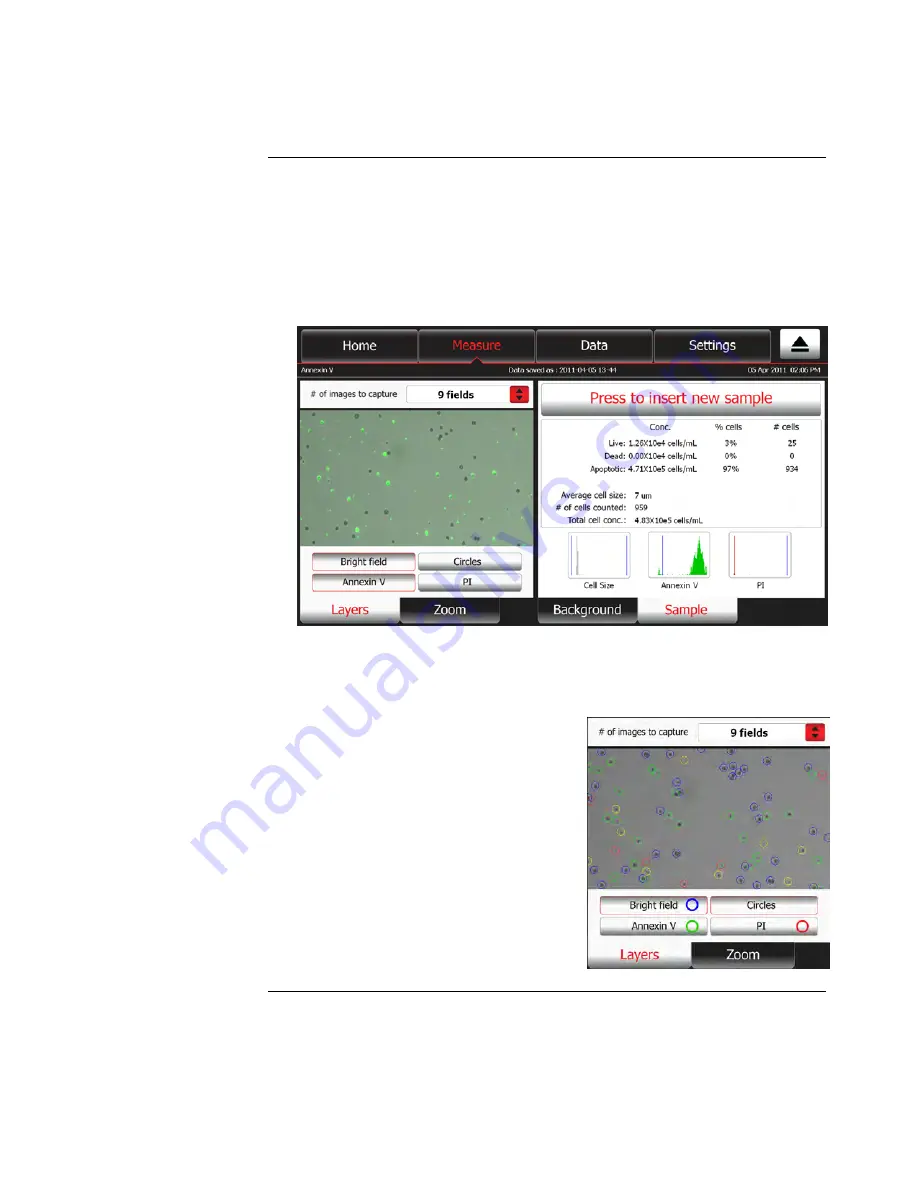
To review images captured through different channels (bright field, green
fluorescence, red fluorescence), select the image by touching the
thumbnail
in
the
Zoom tab
, and then press the
Layers tab
.
Note:
The number of layers that are available for viewing and the layer button labels
depend on the Tali
™
Assay performed. For example, the button for the green-fluorescent
channel is labeled Annexin V and the button for the red-fluorescent channel is labeled PI
when the Tali
™
Apoptosis Assay is selected (see image below), while the same buttons
would be labeled SYTOX Blue and RFP if the RFP + Viability assay was selected.
2.
Touch appropriate button on the Layers tab to display the image captured in a
particular channel (in this example, touch
Annexin V
or
PI
). Press the same
button again to remove the layer from the display.
Note:
You may superimpose layers by touching multiple layer buttons
3.
To identify the cells counted in a
particular channel, touch
Circles
.
The Tali
™
Image-Based Cytometer circles
the cells that were analyzed as follows:
Blue:
cells counted in bright field
channel
Green:
cells counted in green-
fluorescence channel
Red:
cells counted in red-
fluorescence channel
Yellow:
cells counted in both green
and red channels
Black:
cells excluded from the count
Continued on next page


