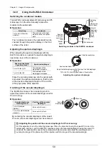
background image

Chapter 3    Usage of Components 

41 

  3.4    Using the Condenser Section 

  3.4.1    Selecting a Suitable Condenser 

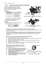
The following four types of condensers can be used with this microscope:   
Select a condenser suitable for microscopy. 


Condenser turret (system condenser) 

Up to seven condenser modules can be attached. 
Microscopy techniques can be switched by turning 
the turret to change the condenser module. 

The condenser lens is replaceable and a range of 
lenses is available for the applicable microscopy. 


Condenser turret (system condenser) 


ELWD-S condenser 

This condenser allows BF microscopy and Ph 
microscopy. Manually turn the turret to select optical 
elements. 

Note:  An ELWD condenser lens, which cannot be 

replaced, is mounted on the ELWD-S 
condenser.  


ELWD-S condenser 


HNA condenser slider 

A high-magnification DIC or Ph microscopy (or BF 
microscopy) is available in combination with a high 
NA condenser lens. Manually pull in or pull out the 
slider using the slider in/out levers to select a 
condenser module. 

Two types of high NA condenser lenses can be used: 

• 

TI2-C-LHD HNA dry lens 

• 

TI2-C-LHO HNA oil lens 

Note:  A high NA lens is required for using the HNA 

condenser slider. 


HNA condenser slider 


DF condenser 

This is a condenser exclusively for DF microscopy. 
There are two types of condensers and mounted 
immediately under the TI-DF condenser adapter. 

• 

Darkfield condenser oil 

• 

Darkfield condenser dry 


DF condenser 


Condenser lens

Turret

Centering knob

clamp screw

(both sides)

Condenser

centering screw

(both sides)

Aperture diaphragm 

lever 

DF condenser 

TI-DF Dark-Field 

Condenser 

Adapter 

Extension tube 

(supplied with the 

high NA lens) 

Slider in/out lever 

(both sides) 

Slider 

Aperture

diaphragm lever

Dust-proof

plate

High NA condenser lens 

Aperture 

diaphragm lever

Condenser lens 

Condenser

module indication

Condenser module 

replacement port 

Turret 

Module centering

screws

(both sides)

Содержание ECLIPSE T-17-369

Страница 1: ...M702E 21 2 NF 8 1 2 M702EN08 Inverted Research Microscope Instruction Manual ...

Страница 2: ......

Страница 3: ...actual product in its appearance Although every effort has been made to ensure the accuracy of this manual errors or inconsistencies might remain If you notice any points that are unclear or incorrect please contact your local Nikon representative Some of the equipment described in this manual may not be included in the set you have purchased If you intend to use any other equipment with this prod...

Страница 4: ... 3 2 Right and Left Operation Panels 12 1 3 3 Power Switch 13 1 3 4 AC Adapter and Power Cord 13 Chapter 2 Microscopy 15 2 1 Diascopic Bright field BF Microscopy 16 2 2 Diascopic Phase Contrast Ph Microscopy 18 2 3 Diascopic DIC IMSI Microscopy 20 2 4 Episcopic Fluorescence Epi FL Microscopy 22 2 5 Diascopic Dark field DF Microscopy 24 2 6 NAMC Microscopy 26 Chapter 3 Usage of Components 29 3 1 Po...

Страница 5: ...e Unit with Port 57 3 8 Using the Stage 58 3 8 1 Setting a Sample on the Manual Stage 58 3 8 2 Setting a Sample on the Plain Stage Gliding Stage 59 3 8 3 Moving a Sample on the Stage 60 3 9 Using the Nosepiece 61 3 9 1 Turning the Nosepiece to Switch the Objective 61 3 9 2 Placing the DIC Prism into the Optical Path 61 3 10 Using the Objective 62 3 10 1 Adjusting the Objective with the Correction ...

Страница 6: ...rinciples of DF Microscopy 97 4 5 2 Optical Elements Required for DF Microscopy 97 4 5 3 Combination of the Optical Elements Required for DF Microscopy 99 4 6 Details of NAMC Microscopy 100 4 6 1 Principles of NAMC Microscopy 100 4 6 2 Optical Elements Required for NAMC Microscopy 101 4 6 3 Combination of Optical Elements 102 Chapter 5 Troubleshooting 103 5 1 Troubleshooting for Microscopy Techniq...

Страница 7: ... 144 7 8 3 Attaching a Fixed Filter for Dia illumination 145 7 8 4 Attaching a Filter Slider for Dia Illumination Optional 146 7 8 5 Attaching a Motorized Dia Illumination Shutter Optional 147 7 8 6 Mounting a Lamphouse for Dia illumination 148 7 8 7 Replacing a Dia Illumination Lamp Precentered Lamphouse only 149 7 9 Attaching a Contrast Shield Optional 150 7 10 Attaching an Analyzer and a Polari...

Страница 8: ...6 2 Parfocal Adjustment of Objectives without Correction Collar 173 7 17 Attaching a Specimen Holder 175 7 17 1 Attaching a Specimen Holder for the Manual Stage 175 7 17 2 Attaching a Stage Ring 175 7 18 Moving the Microscope 176 Chapter 8 Specifications 177 8 1 Principles of Operation 178 8 2 Performance Properties 179 8 3 Physical Properties 183 ...

Страница 9: ...Manual stage Plain stage Gliding stage Caution for heat This symbol label is attached on the top surface of the optical path cover on the top of the illumination pillar on the top surface of the TI2 D SF filter slider for dia illumination and on the back surface of the D LH LC precentered lamphouse When the D LH LC is used as the dia illumination source this symbol reminds the user of the followin...

Страница 10: ... cube replacement port of the FL turret to remind the user of the following precautions Be sure to close the cover of the filter cube replacement port because light including ultraviolet light might leak from the port For details see 12 Cautions on replacing the filter cube under WARNING Close the shutter before opening the cover of the filter cube slot C FL turret WARNING and CAUTION Symbols Used...

Страница 11: ...cause malfunction or fire This product is classified into class 1 in terms of protection against electric shock and must therefore be connected to a protective grounding terminal For details on the specified power cord see Chapter 8 Specifications To protect from electric shock always turn off the power switch set it to o before connecting power cords 5 Use the specified lamps Be sure to use only ...

Страница 12: ...immersion oil for oil immersion lenses Absolute alcohol ethyl alcohol or methyl alcohol for cleaning optical components Petroleum benzine for removing immersion oil Medical alcohol for disinfecting the microscope Keep these solvents away from fire Before using a solvent carefully read the instructions provid ed by the manufacturer of the solvent and handle it correctly and safely Note the followin...

Страница 13: ...epi illumination section when the risk group classification is the Exempt Group which does not cause photobiological hazards is shown below This table shows the highest risk when a D LEDI fluorescence LED illumination system is used as a light source Risk group classification Hazard distance UV hazard Risk group 3 0 9 m or more Retinal blue light hazard Risk group 2 4 1 m or more The emitted light...

Страница 14: ...sk Group 1 IR light is emitted from the aperture of the condenser lens when dia illumination is used Use appropriate shielding or eye protection Precentered lamphouse Caution labels indicating photobiological safety are affixed on the D LH LC precentered lam phouse for dia illumination to remind the user of the precaution below CAUTION UV emitted from this product Eye or skin irritation may result...

Страница 15: ...e shutter before opening the cover of the filter cube slot Caution label fluorescence filter cube replacement port cover of the FL turret Be sure to close the shutter of the FL turret when opening the filter cube replacement port If the cover is open illumination including ultraviolet light will leak from the aperture when the cover of the replacement port is opened possibly disturbing epi fl micr...

Страница 16: ...the top of this product In particular never place a heavy object on top of the microscope as it might cause deformation damage or malfunction of the product or injury if the object falls 4 Do not turn on the power when this product is covered Do not turn on the power when this product is covered by anything Doing so might block ventila tion of the microscope and cause it to overheat possibly resul...

Страница 17: ...amps Dispose of used lamps according to the regulations or rules of your local government or ask a licensed industrial waste disposal operator to dispose of it 10 Cautions on carrying the microscope When carrying the microscope main body observe the following precautions to ensure safety Failure to observe these precautions might cause the microscope to drop possibly resulting in damage or serious...

Страница 18: ... to hold the specified positions when carry ing the main body When carrying only the microscope main body base with all the devices removed firmly hold the microscope by gripping the front bottom and rear recesses of the main body 11 Cautions on assembly and replacement Take care not to pinch your fingers or hands in a joint or under a movable part Scratches and dirt such as fingerprints on optica...

Страница 19: ...e 4 Installation location and storage location This product is a precision optical instrument Use or storage of this product under inappropriate conditions might result in malfunction or loss of precision For details on the temperature and humidity at the installation location and storage location see 8 3 Physical Properties The following conditions must also be considered Install and store this p...

Страница 20: ...ery of the stage thereby affecting the microscopy work After opening an immersion oil container use oil as early as possible because oil deteriorates and its refractive index and viscosity change When using petroleum benzine or absolute alcohol to wipe off immersion oil or to clean the lenses follow the instructions provided by the manufacturer of the petroleum benzine or alcohol to be used Keep t...

Страница 21: ... Their Functions This chapter describes the name and function of each unit and operation section of the microscope system in a basic configuration For details on how to use each component see Chapter 3 Usage of Components For details on the components of the entire microscope system see Chapter 7 Assembly of the Devices ...

Страница 22: ...ion Tube base unit Condenser turret Nosepiece FL turret Epi fluorescence attachment or back port optional Manual stage Stage holder Shielding plate Polarizer Side port right Front operation panel microscope Analyzer slider Objective Right operation panel microscope D LEDI fluorescence LED illumination system with a remote control pad Photobiological safety label Eyepieces Left operation panel micr...

Страница 23: ...age TC S SR Stage handle Long handle TI2 S HL Stage holder Ring holder set C S HLS Nosepiece DIC sextuple nosepiece TI2 N ND Objective CFI objective Port Direct C mount adapter Diascopic illumination lamphouse Diascopic LED lamphouse TI2 D LHLED Pillar for dia illumination Pillar for dia illumination TI2 D PD Condenser Condenser turret TC C TC LWD condenser lens TI C LWD Polarizer DIC polarizer TI...

Страница 24: ... the LED of the dia illumination section is prevented from emitting autofluorescence due to epi illumination Fixed filter slot Up to 2 filters can be inserted 3 3 4 Lamphouse for Dia illumination Control this lamphouse by the dia illumination ON OFF button and the light control volume 3 3 2 and 3 3 7 Dia illumination shutter knob C Close Close the shutter O Open Open the shutter 3 3 1 Field diaphr...

Страница 25: ...wer limit 3 4 2 Condenser turret Turning the turret switches the condenser module Through the front window the address of the module that enters the optical path can be checked 3 4 6 Condenser centering screws Adjust the position of the condenser 3 4 3 Condenser lens 3 4 Polarizer Change dia illumination to polarized illumination 3 5 1 Polarizer slider Use this slider to switch between the polariz...

Страница 26: ...inocular part The distance between the eyepieces can be adjusted according to the interpupillary distance of the user 3 6 3 Angle adjustment mechanism ER tube The angle of the binocular part can be adjusted in the range of 15 to 45 degrees 3 6 1 Eyepiece Diopter can be adjusted according to the observer s eyesight 3 6 2 Ultraviolet light shielding plate This plate prevents ultraviolet light or str...

Страница 27: ... handle 3 8 3 Holder mount The user can mount a specimen holder suitable for the specimen to be used This figure shows a universal holder mounted 3 8 1 Stage handle Use this handle to move the stage Upper knob Move the stage back and forth Lower knob Move the stage to the right and left 3 8 3 The stage handle can be selected from three types long handle middle handle and short handle ...

Страница 28: ...prism N1 N2 and NR NAMC objective Indicates a NAMC code NAMC1 to NAMC3 Nosepiece When switching the objective turn the turret on the nosepiece Turning the turret clockwise Move to the next address on the nosepiece Turning the turret counterclockwise Move back to the previous address on the nosepiece 3 9 1 Objective 3 10 DIC slider slot Insert the DIC slider in this slot for DIC microscopy 3 9 2 Co...

Страница 29: ...he FL turret to place the analyzer into the optical path Analyzer block Polarizing plate Filter cube replacement port Attach the filter cube to the inside through this port The plate holder indicating the type of the attached filter cube is provided Analyzer slider 3 5 2 Turret address indication This indicates the address of the filter cube currently in the optical path The address is displayed o...

Страница 30: ...rear view Right front view Fixed main branch EPI FL module Epi fluorescence attachment Illumination module EPI FL module Field diaphragm slider Push the slider in until it clicks into position The field diaphragm enters the optical path Pull the slider out until it reaches the limit position The field diaphragm is removed from the optical path 3 13 3 Aperture diaphragm centering knobs x 2 one on t...

Страница 31: ...n system controls respective brightness of four types of LEDs of different wavelengths by turning on or off those LEDs respectively A dedicated remote control pad is used to control the fluorescence LED light source PUSH ON OFF SELECT Fn Manu facturer NIKO N CORPORATION Shina gawa Interc ity Towe r C 2 15 3 Konan Minato ku Tokyo 108 6 290 Japan Importer in the European Economic Area EC REP NIKON I...

Страница 32: ...o vertically move the objective focusing device in coarse mode 3 11 1 Dia illumination ON OFF button Use this button to turn on off the dia illumination 3 3 2 Dia illumination brightness adjuster Use this knob to adjust brightness of the dia illumination 3 3 7 Refocusing lever Use this lever to set the current position of the coarse focus knob as the upper limit position 3 11 3 Pushing this lever ...

Страница 33: ...itch for the Ti2 U main body 1 3 4 AC Adapter and Power Cord The AC adapter and the power cord are used to supply power to the Ti2 U main body AC adapter and power cord About the AC adapter and power cord Be sure to use the AC adapter supplied with this product For details on the AC adapter see Chapter 8 Specifications Be sure to use only the power cord specified in Chapter 8 Specifications when c...

Страница 34: ...Chapter 1 Names of Parts and Their Functions 14 This page is intentionally blank ...

Страница 35: ... 6 System configuration The microscopy procedure is described based on a microscopy system in the basic configuration described in Chapter 1 Names of Parts and Their Functions If any of these microscopy techniques require additional parts or components they are described on the pages corresponding to such techniques Setup of the system The following procedures assume that all the necessary devices...

Страница 36: ...e the FL turret shutter h Dia illumination shutter knob 3 3 1 i FL turret shutter knob 3 12 2 2 2 Turn on the dia illumination LED and adjust the brightness j Dia illumination ON OFF button 3 3 2 k Dia illumination brightness adjuster 3 3 2 Illumination reaches the observation port Use filters if required 2 3 Fully open the dia field diaphragm l Field diaphragm dial 3 3 3 2 4 Fully open the dia ap...

Страница 37: ...the objective to the objective with a specified magnification b Nosepiece 3 9 1 4 2 When using a liquid immersion type objective perform oil or water immersion g Objective 3 10 2 and 3 10 3 4 3 Remove one of the eyepieces attach the centering telescope and focus on the pupil of the objective r Centering telescope 3 6 4 4 4 Adjust the size of the dia aperture diaphragm to 70 80 of the size of the n...

Страница 38: ...adjust the phase plate ring and annular diaphragm 2 1 Change the objective to the objective for Ph microscopy a Nosepiece 3 9 1 The phase plate ring enters the optical path Memorize the Ph code of the objective 2 2 When using a liquid immersion type objective perform oil or water immersion b Objective 3 10 2 3 10 3 2 3 Focus on the sample c Focus knob 3 11 1 2 4 Place the Ph condenser module into ...

Страница 39: ...ield diaphragm dial 3 3 3 3 3 When using an objective with a correction collar adjust the correction collar b Objective 3 10 1 Adjust the correction collar of the objective according to the thickness of the prepared specimen slide or the cover glass of the culture vessel 3 4 Move the stage and turn the focus knobs to observe the sample i Stage handle 3 8 3 c Focus knob 3 11 1 3 5 When using anothe...

Страница 40: ...g plate 2 1 Change the objective to the objective for DIC or IMSI microscopy a Nosepiece 3 9 1 2 2 When using a liquid immersion type objective perform oil or water immersion b Objective 3 10 2 3 10 3 2 3 Focus on the sample c Focus knob 3 11 1 2 4 Move the field of view to the portion of the specimen where no object is visible or the portion of a prepared specimen slide where a cover glass is pla...

Страница 41: ...1 3 3 Remove one of the eyepieces attach the centering telescope and focus on the pupil of the objective e Centering telescope 3 6 4 3 4 Adjust the size of the dia aperture diaphragm to 70 80 of the size of the numerical aper ture of the objective j Aperture diaphragm open close lever 3 4 5 Adjust the size of the aperture diaphragm image while observing the objective pupil plane and the aperture d...

Страница 42: ...tofluorescence due to epi illumination close the dia illumination shutter g Dia illumination shutter knob 3 3 1 h Condenser turret 3 4 6 2 2 Specify the FL turret address and place the filter cube suitable for the sample into the optical path i FL turret 3 12 1 2 3 Fully open the epi aperture diaphragm j Aperture diaphragm open close lever 3 13 1 2 4 Place the field diaphragm slider into the optic...

Страница 43: ...cedure from step 3 1 to step 3 3 4 Perform Epi FL microscopy 4 1 Change the objective to the objective for fluorescence microscopy with a specified magnification b Nosepiece 3 9 1 4 2 When using a liquid immersion type objective perform oil or water immersion f Objective 3 10 2 3 10 3 4 3 Adjust the size of the epi field diaphragm image and move it to the center of the field of view k Field diaphr...

Страница 44: ...shutter and close the FL turret shutter g Dia illumination shutter knob 3 3 1 h FL turret shutter knob 3 12 2 2 2 Turn on the dia illumination LED and adjust the brightness i Dia illumination ON OFF button 3 3 2 j Dia illumination brightness adjuster 3 3 2 Illumination reaches the observation port Use filters if required 2 3 Fully open the dia field diaphragm k Field diaphragm dial 3 3 3 2 4 Place...

Страница 45: ... a specified magnification b Nosepiece 3 9 1 4 2 When using a liquid immersion type objective perform oil or water immersion f Objective 3 10 2 3 10 3 4 3 When using an objective with a diaphragm align the diaphragm to an optimum position below the minimum numerical aperture of the condenser f Objective 3 10 4 4 4 When an objective with correction collar is used adjust the correction collar f Obje...

Страница 46: ... a Nosepiece 3 9 1 2 2 Focus on the sample b Focus knob 3 11 1 2 3 Place the NAMC condenser module having the same NAMC code as the objective into the optical path c Condenser turret 3 4 6 2 4 Remove one of the eyepieces and attach the centering telescope d Centering telescope 3 6 4 2 5 Check that the polarizer for NAMC IMSI is in the optical path e Polarizer for NAMC IMSI 3 5 1 2 6 Turn the eyepi...

Страница 47: ...telescope 3 6 4 3 Perform NAMC microscopy 3 1 Fully open the dia aperture diaphragm h Aperture diaphragm open close lever 3 4 5 3 2 Adjust the size of the dia field diaphragm image i Field diaphragm dial 3 3 3 3 3 When using an objective with a correction collar adjust the correction collar f Objective 3 10 1 Adjust the correction collar of the objective according to the thickness of the prepared ...

Страница 48: ...Chapter 2 Microscopy 28 This page is intentionally blank ...

Страница 49: ...se each module and device in the Ti2 U main body CAUTION Before using the product thoroughly read the Safety Precautions at the beginning of this manual and heed all warnings and cautions written therein To use other products together with this product refer to the respective manuals and heed all warnings and cautions written therein ...

Страница 50: ...dia illumination the dia illumination can be controlled from the Ti2 U main body by connecting the Ti2 U main body to the power supply for dia illumination When controlling the power supply from the microscope main body make sure that the Ti2 U main body and the power supply are correctly connected and that the external control for the power supply is set to ON 1 Ti2 U microscope main body 2 Perip...

Страница 51: ...ime of purchase There is no output if not specified R80 Right side port 80 Eyepiece observation port 20 Ti2 U output port The following output ports are usable in this microscope Output port Description Eyepiece observation port An optical output port for visually observing a microscope image through a binocular part of a tube Side port right left An optical output port for a microscope image on b...

Страница 52: ...e Turn in the opposite direction of the arrow vertical upward 1x Turn in the direction of the arrow until 1 5x aligns with the index mark sideways 1 5x 1 5x Device magnification changeover Using the intermediate magnification dial The settings of the intermediate magnification dial are valid for all optical image output ports except the optional back port By replacing the 2nd tube lens inside the ...

Страница 53: ...scence due to epi illumination 3 3 2 Using Diascopic Illumination Control dia illumination using the dia illumination ON OFF button and brightness adjuster on the left side of the microscope Operation Turning on off dia illumination Dia illumination ON OFF button Dia illumination Press the button Pressing the button alternates between on and off Adjusting dia illumination Brightness adjuster Dia i...

Страница 54: ...the optical image decreases Procedure for adjusting the field diaphragm 1 Place the objective with a specified magnification into the optical path 2 Turn the field diaphragm dial to stop down the field diaphragm until the field diaphragm image is seen in the field of view 3 Turn the condenser focus knob to focus on the field diaphragm image 4 Turn the condenser centering screw to move the field di...

Страница 55: ...mination each with two filters two fixed filters can be mounted and up to four filters can be placed into the optical path simultaneously Usable filters Filter name Explanation ND2 ND4 ND8 ND16 Light absorbing filter ND Neutral Density for adjusting brightness The greater the number the lower the transmittance and the darker the image becomes This filter does not affect the color shade of the illu...

Страница 56: ...pillar to prevent the pillar from falling unintentionally even though the pillar is usable with the clamp released When a motorized shutter or precentered lamphouse for dia illumination is attached in particular make sure to use the pillar with the pillar tilting clamp knob tightened Attach a third party part to the illumination pillar There are eight M4 screw holes on the front and the side of th...

Страница 57: ...ght shielding plate Light is not blocked Close the light shielding plate Light is blocked S F Operating the contrast shield Mounting position of the contrast shield The contrast shied can be mounted at two positions 60 mm or 30 mm from the stage surface The shorter the distance from the stage surface the higher the light shielding performance but it might become more difficult to manipulate the sp...

Страница 58: ...of a lamphouse and connection of a power supply For details on the attachment of a lamphouse and connection of a power supply see Chapter 7 Assembly of the Devices Checking the settings of the power supply When turning on or off or adjusting brightness of the lamp from the main body check the following settings 1 Check the connection between the power supply and the Ti2 U main body 2 Set the EXTER...

Страница 59: ...ER indicator lights The brightness can be checked by the index of the brightness adjuster Operation panel of the power supply The dia illumination ON OFF button on the left side of the microscope is enabled even when the EXTERNAL switch is turned off Before adjusting the brightness of the lamp from the power supply turn the brightness adjuster on the left side of the microscope counterclockwise so...

Страница 60: ...entered lamphouse and motorized shutter Operating the motorized shutter controller The motorized shutter controller is equipped with two operation switches SHUTTER 1 and SHUTTER 2 which enable two motorized shutters to be controlled by a single controller Operation Opening and closing the shutter SHUTTER 1 switch SHUTTER 2 switch Motorized shutter Press the switch The shutter opens or closes each ...

Страница 61: ...copy is available in combination with a high NA condenser lens Manually pull in or pull out the slider using the slider in out levers to select a condenser module Two types of high NA condenser lenses can be used TI2 C LHD HNA dry lens TI2 C LHO HNA oil lens Note A high NA lens is required for using the HNA condenser slider HNA condenser slider DF condenser This is a condenser exclusively for DF m...

Страница 62: ...use the condenser to slide down by its own weight Using the condenser refocusing clamp Even when the condenser is moved upward to the escape position to replace samples the condenser can be returned to the original position by lowering to the limit position Use this clamp when using a high NA condenser or when the dia illumination pillar cannot be tilted 3 4 3 Adjusting the Position of the Condens...

Страница 63: ... the condenser refocusing clamp After forming the field aperture image on the specimen plane tighten the condenser refocusing clamp and mark the power limit of the condenser This prevents the tip of the condenser lens from colliding with the specimen and affixes the field aperture image Wiping off oil Oil remaining at the tip of the oil immersion lens after the wiping or oil adhering to the tip of...

Страница 64: ...just the aperture diaphragm size according to specimens to be observed and the purpose of the observation Timing of aperture diaphragm adjustment and tips on adjustment Adjust the field diaphragm every time the objective is switched Generally images with an appropriate contrast can be obtained by narrowing the field aperture to 70 to 80 of the numerical aperture of the objective Adjustment of the ...

Страница 65: ...r The DIC module enters the optical path Pull it out to the front The Ph module or hollow enters the optical path Switching modules by the HNA condenser slider When switching the condenser module manually turn the turret ring to place the module into the optical path Operation Turret ring Condenser turret Turn until it clicks into position The front or rear module enters the optical path The addre...

Страница 66: ...C Module TI2 C MH N2D High NA Dry DIC Module TI2 C MH NRD High NA Dry DIC Module TI2 C MH N2O High NA Dry DIC Module TI2 C MH NRO High NA Dry DIC Module NAMC NAMC TC C MN 10x NAMC 10x Module TC C MN 20x NAMC 20x Module TC C MN 40x NAMC 40x Module TC C MNL 10x NAMC LWD 10x Module TC C MNL 20x NAMC LWD 20x Module TC C MNL 40x NAMC LWD 40x Module IMSI IMSI TC C MI N2D IMSI Dry Module TC C MI NRD IMSI...

Страница 67: ...ring diaphragm image by turning the eyepiece section of the centering telescope Turn the centering screws of the module using a specified tool to overlay the annular diaphragm image on the phase ring image in the objective Condenser turret HNA condenser slider Annular diaphragm centering screw Centering of the annular diaphragm Phase ring Annular diaphragm Annular diaphragm centering screws Annula...

Страница 68: ...e orientation of the modulator according to the specimen Modulator adjustment The modulator image can be observed using the centering telescope Turn the modulator ring and adjust the orientation of the modulator so that contrast can be seen in the required direction Operation Operation Polarizing plate Turn the modulator ring The orientation of the modulator is changed Modulator image Adjustment b...

Страница 69: ...y zone G Operation Module adjustment Polarizing plate Turn the centering screw The position of the slit diaphragm is changed Turn the module through the module rotation slot The orientation of the slit diaphragm is changed Slit diaphragm image Slit diaphragm adjustment Fine adjustment of the NAMC condenser slit diaphragms Images obtained by NAMC microscopy vary depending on the slit diaphragm adju...

Страница 70: ...he field diaphragm so that the size of the field diaphragm image is 70 to 80 that of the pupil plane of the objective Adjusting the aperture diaphragm Centering of the annular diaphragm The ELWD S condenser has a centering knob to adjust the position of the annular diaphragm used for Ph microscopy Operation Annular diaphragm centering screw Position of the annular diaphragm Turn it clockwise tight...

Страница 71: ...ow hole Placing the polarization plate in the optical path will change the illumination from dia illumination to polarized illumination Inserting and removing the polarizer NAMC IMSI polarizer The NAMC IMSI polarizer has no slider When placing the polarizer into the optical path attach the polarizer to the condenser mount When removing the polarizer from the optical path remove the polarizer from ...

Страница 72: ...9 Remove one of the eyepieces and attach the centering telescope 10 Turn the entire polarizer until a dark cross appears 11 With the dark cross in view tighten the clamp screw using a hexagonal screwdriver so that the polarizer is fixed in place 12 Replace the centering telescope with the removed eyepiece Attaching a polarizer Adjusting and fixing the polarizer Dark cross on the pupil plane If no ...

Страница 73: ...nalyzer slider van be inserted from either the right or left side of the microscope main body Close the shutter before opening the cover of the filter cube slot C Inserting and removing the analyzer slider Placing the analyzer block in the optical path Operation FL turret Analyzer Place the address where the analyzer block is attached into the optical path The analyzer enters the optical path Set ...

Страница 74: ...Binocular part Angle of the binocular part Turn the binocular part upward The eyepiece faces upward max 45 degrees Turn the binocular part downward The azimuth of the eyepiece moves closer to horizontal min 15 degrees Adjust the angle according to your preference for comfortable viewing Adjusting the angle of the binocular part In the case of using an S tube The S tube TC T TS has no angle adjustm...

Страница 75: ...nce between the right and left eyes are corrected so that observation with both eyes is performed easily 1 In the BF microscopy status focus the 10x objective on the specimen 2 Turn the diopter correction collar of the eyepiece for each eye to align the ruling line of the diopter correction collar with the rim of the outer ring of the eyepiece This position is used as the position of diopter corre...

Страница 76: ...he pupillary distance is helpful so that pupillary distance can be easily adjusted the next time 3 6 4 Operating the Centering Telescope The centering telescope is an optical instrument that is mounted on the binocular part to observe the pupil plane of the objective The centering telescope is used for different purposes such as adjusting the aperture diaphragm checking for air bubbles during oil ...

Страница 77: ...r knob Operation Optical path changeover knob Optical path of the tube base unit EYE Eyepiece observation port CAMERA Tube base unit side port Switching the optical path of the tube base unit Switching the optical path To output microscope images from the side port of the tube base unit select EYE using the optical path changeover knob of the microscope main body In the case of using an S tube bas...

Страница 78: ...upper surface of the stage there are screw holes for fixing an auxiliary module such as a manipulator When using a laser light source attach a laser safety cover This section explains how to use the universal holder petridish holder and well clamper Using the universal holder The universal holder is used to hold a glass slide prepared specimen slide or dish in place Operation Adjust the width of t...

Страница 79: ... it on the holder section of the stage Close the well clamper gently and fix the well plate Using the well clamper 3 8 2 Setting a Sample on the Plain Stage Gliding Stage Various types of stage rings can be mounted on the TI SP Plain Stage and the TC S GR Gliding Stage Operation Put a petri dish or a glass slide prepared specimen slide on the stage ring When using a gliding stage use an optional s...

Страница 80: ... details on the procedure for setting the stroke see Chapter 7 Assembly of the Devices Rack protection plate for the manual stage A protection plate for protecting the rack of the stage is located on the top of the stage handle Because the protection plate protrudes from the stage take due care not to bump your hands or other objects against the plate Using a gliding stage By moving the movable pa...

Страница 81: ...ctive by turning the nosepiece move the focusing device to lower the objective and then turn the nosepiece so as to prevent the objective from contacting the specimen 3 11 3 9 2 Placing the DIC Prism into the Optical Path When performing DIC microscopy insert the DIC slider in the DIC slider slot of the nosepiece Operation DIC slider DIC prism Press in the slider up to the limit position The DIC p...

Страница 82: ...the specimen with the focus knobs 3 If the resolution and contrast of the image are poor slightly turn the correction collar on the objective in either direction This will shift the focus slightly Readjust the focus by turning the fine focus knob 4 If the image becomes clearer slightly turn the correction collar again in the same direction If the image becomes less clear slightly turn the collar i...

Страница 83: ...use a cover glass with a thickness of 0 17 mm In this case face the cover glass downward and set the specimen so that the cover glass faces the objective Glass thickness of 1 2 mm When using the objective labeled 1 2 use a normal cover glass with a thickness of 1 2 mm In this case face the cover glass upward and set the specimen so that the glass slide faces the objective Glass thickness of 0 17 m...

Страница 84: ... stage and the focus knobs 7 After using an oil immersion objective wipe the oil off the tip Oil immersion Handling of immersion oil If air bubbles are mixed in the immersion oil the quality of the microscope images will decrease When using immersion oil take care not to let air bubbles get mixed in Oil remaining at the tip of the oil immersion lens after the wiping or oil adhering to the tip of t...

Страница 85: ...After using a water immersion objective wipe the water off the tip Example of a water immersion objective Handling a water immersion objective When immersing an objective in water avoid using tap water There are impurities in tap water that might adhere to the glass surface and harden when the water at the lens tip dries off This might cause the lens to be scratched or damaged at the time of clean...

Страница 86: ...of the objective according to the NA of the DF condenser Operation Check the minimum NA of the DF condenser and then adjust the diaphragm adjusting ring so that the NA of the objective is smaller than the minimum NA Display Check the numerical aperture using the gradua tions on the diaphragm adjusting ring Example of an objective with a diaphragm Iris mark Diaphragm adjusting ring ...

Страница 87: ...sing device moves downward in coarse motion mode Degree of objective movement The following table shows the degree of objective movement when the focus knobs are turned Degree of knob rotation Degree of objective movement One graduation step of the fine focus knob 1 μm One turn of the fine focus knob 0 1 mm One turn of the coarse focus knob 5 mm Close the shutter before opening the cover of the fi...

Страница 88: ...ing the torque of the coarse focus knob Operation Coarse focus knob torque adjustment ring Torque of the coarse focus knob Turn the ring to the rear counterclockwise Increase Turn the ring to the front clockwise Decrease Note The torque of the fine focus knob is fixed Using the coarse focus knob torque adjustment ring Adjusting the coarse focus knob torque Excessively loosening the coarse focus kn...

Страница 89: ...tion of the CLAMP arrow The knob is restricted to prevent the objective from moving beyond the current position Pull the lever to the front in the opposite direction to the CLAMP arrow Vertical movement restriction is released This lever does not influence the movement of the fine focus knob This means that the objective can be moved above the limit position by turning the fine focus knob Using th...

Страница 90: ...er inside the FL turret can be opened and closed using the shutter lever Operation Shutter lever FL turret shutter Move the lever to the O side front The shutter opens Move the lever to the C side rear The shutter closes Close the shutter before opening the cover of the filter cube slot C Opening and closing the FL turret shutter Use of FL turrets in a two tier configuration FL turrets can be used...

Страница 91: ...hen a camera with a field number of 22 mm on the imaging surface is used Conventional filter cube uses a 25 mm filter Applicable microscopy The thick dichroic mirror is suitable for applications that require high degree of the dichroic mirror flatness such as TIRF observation Maintaining mounting compatibility The mount part for the FL turret is compatible with the conventional 25 mm diameter seri...

Страница 92: ...ragm decreases The narrower the aperture diaphragm is the smaller the amount of stray light becomes but the darker the image of the specimen becomes The wider the aperture diaphragm is the brighter the image becomes but the larger the amount of stray light becomes By narrowing the aperture diaphragm to the size equal to the pupil diameter of the objective brightness of the specimen image can be ke...

Страница 93: ...hich allows specific wavelengths of light from the light source to pass through is intended for Epi FL microscopy Normally the excitation filter is used by attaching a filter cube to the FL turret By attaching an excitation filter to the filter slider of the epi fluorescence attachment the excitation filter can be switched separately from the filter cube Combination of an excitation filter and a b...

Страница 94: ...hragm slider during large field of view microscopy To perform microscopy with a large field of view with a field number of equivalent to or more than 22 remove the field diaphragm slider from the optical path Adjusting the size of the field diaphragm Adjust the size of the field diaphragm using the field diaphragm open close lever of the field diaphragm slider Operation Field diaphragm open close ...

Страница 95: ...m image to the center of the field again Field diaphragm image Eyepiece field ring Centering and size adjustment for the field diaphragm Differences by diaphragm shape There are three types of field diaphragm sliders Each type differs in diaphragm shape and has the following characteristics Circular field stop slider TI2 F FSC Slider with a circular field diaphragm Square field stop slider TI2 F F...

Страница 96: ...status of the optical path of the main branch can be identified as the inser tion removal status of the optical path changeover lever Switching the optical path of the main branch Using the optical path changeover lever Using the switching mirror The optical path of the main branch can be switched by inserting or removing the internal switching mirror Although only full mirrors and half mirrors ar...

Страница 97: ... a back port unit Focusing Loosening the clamp screws allows turning the focus adjustment knob Turn the focus adjustment knob while watching a camera captured image to align it with an observation image in the eyepieces Centering Tightening and loosening two centering screws allows centering a captured image When two cameras are used for a simultaneous capture make sure that the images captured by...

Страница 98: ...by the camera so that the view of the image becomes equivalent to the image observed through the eyepieces After focus adjustment is complete tighten the focus adjustment fixing screw to fix the focus adjustment section Centering adjustment for the camera device Tightening and loosening two centering screws allows centering a captured image Adjustment tool When rotating the focus adjustment fixing...

Страница 99: ...iques 79 Chapter 4 Microscopy Techniques Chapter 4 Microscopy Techniques This chapter explains the principles of each microscopy and optical elements required for implementing each microscopy and combinations of optical elements ...

Страница 100: ...he illumination also changes Because of this incidence angle θ adjustment of the aperture diaphragm causes the contrast resolution and focal depth of images to change Use of fly eye lens to make illumination uniform The uniformity of the illumination can be further increased using fly eye lenses for the illumination attachment A fly eye lens is an array of small hexagonal lenses The name fly eye l...

Страница 101: ...opy BF microscopy is not suitable for observing colorless transparent specimens that do not absorb light 4 1 2 Optical Elements Required for BF Microscopy Dia illumination unit TI2 D PD dia illumination pillar is used S F C O TI2 D PD Pillar for Dia Illumination Light source The following two types are usable as a light source for dia illumination TI2 D LHLED LED Lamp House for Dia Illumination D ...

Страница 102: ...a dedicated ELWD condenser Specifications of the ELWD S condenser are as follows NA 0 3 WD 65 mm Notes on the use of the ELWD S condenser When using an ELWD S condenser be sure to decrease the light intensity before switching the turret from the position of PH microscopy to that of BF microscopy Otherwise the field of view rapidly becomes bright 4 1 3 Combination of Optical Elements When using a s...

Страница 103: ... the annular diaphragm It passes the condenser lens and then illuminates the specimen The illumination is divided as direct light solid line which has passed the inside of the specimen and optical path dotted line which has passed phase objects and passes the objective A diffraction phenomenon occurs in a portion with a difference in diffraction ratio Therefore diffraction light contains shape inf...

Страница 104: ...zation phase plate is to provide low contrast with diffracted light weakened by a large structure of diameter 10 μm or more while the contrast of fine structure of diameter 10 μm or less is left as is 4 2 2 Optical Elements Required for Ph Microscopy Ph microscopy requires the following components as well as the status of bright field Ph objective including the phase ring in the figure Optical pat...

Страница 105: ...n with a focus on micro contrast Half tone the use range is wide Phase contrast and absorbing objects stained objects in the low and mid ranges Spores of fungus ordinary living cells thick specimens fungus stained specimens insect eggs fat globules crystals etc ADH Hard tone the use range is relatively narrow Transparent object in the low range Fungus protozoa flagellum fibrin fibril fine granule ...

Страница 106: ... common condenser lens unit is required TE C ELWD S condenser Specifications of the ELWD S condenser are as follows NA 0 3 WD 65 mm 4 2 3 Combination of Optical Elements Combinations of the optical elements for Ph microscopy are listed below Combination of the optical elements for Ph microscopy Objective Ph code Condenser lens Condenser module PhL ELWD condenser lens TC C ME PHL ELWD module LWD co...

Страница 107: ...ed from the light source is converted into linear polarization light that vibrates in only one direction on polarizing plate 1 polarizer and into two beams with their mutually perpendicular polarization planes on Nomarski prism 1 The condenser lens converts the two beams into two parallel beams spaced slightly apart and which then pass through the specimen This separation amount shear amount is le...

Страница 108: ...e shift of the separated beam It requires the use of objectives with less distortion with DIC marking because distortion of polarization deteriorates image quality For specimens that have polarization property such as plastic dishes this microscopy cannot be performed 4 3 2 Optical Elements Required for DIC Microscopy DIC microscopy requires the following components as well as the status of bright...

Страница 109: ... conden ser side DIC prism Condenser lens Condenser lenses that allow DIC IMSI microscopy are as follows LWD condenser lens CLWD condenser lens HNA condenser lens dry HNA condenser lens oil Usable condenser lens LWD condenser lens CLWD condenser lens HNA condenser lens dry HNA condenser lens oil NA 0 52 0 72 0 85 1 35 Working distance 30 mm 13 mm 5 mm 1 9 mm including the glass slide thickness of ...

Страница 110: ...alyzer 4 3 3 Combination of Optical Elements To perform DIC IMSI microscopy use a combination of the following optical elements Referring to the following table combine the DIC condenser module and the DIC slider to match the objective and condenser lens in use In addition to standard combinations there are combinations focusing on high contrast or high resolution Select the best combination as re...

Страница 111: ... N2 Dry white background black text 60x I NR Dry white background black text 60x I R Plan Apo 60XC Plan Apo VC 60X Oil Plan Apo λ 60XC Apo TIRF 60XC Oil Plan Fluor 60XS Oil 60x II 60x II R Plan Apo TIRF 60XC Oil Plan Apo 60XC WI 1 Plan Apo VC 60XC WI Plan Apo λ 60X Oil Plan Fluor 60XC Apo λS 60X Oil 2 Plan Apo VC 60XA C WI 60x IV 60x IV R Plan Apo IR 60XC WI SR Plan Apo IR 60XAC WI 100X Plan Apo V...

Страница 112: ...λS 40XC Sil 40x I 40x I 60X Plan Apo 60XA Oil 60x I N2 Dry green background white text 60x I R 60x I N2 Oil yellow background black text 60x I R Plan Apo 60XC Plan Apo VC 60X Oil Plan Apo λ 60XC Apo TIRF 60XC Oil Plan Fluor 60XS Oil 60x II 60x II R 60x II 60x II R Plan Apo TIRF 60XC Oil Plan Apo 60XC WI 1 Plan Apo VC 60XC WI Plan Apo λ 60X Oil Plan Fluor 60XC Apo λS 60X Oil 2 Plan Apo VC 60XA C WI...

Страница 113: ...I objectives and DIC prisms Objective WD mm Applica ble correction collar mm LWD condenser lens white text Standard combination Focus on the contrast Focus on the resolution Condenser module DIC slider Condenser module DIC slider Plan Apo VC 100X Oil 0 13 None IMSI N2 Dry white background black text 100x I IMSI NR Dry white background black text 100x I R Plan LWD IMSI 100XC 0 95 0 6 1 3 IMSI N2 Dr...

Страница 114: ...optical elements of a filter cube are shown in the following figure 100 80 60 40 20 0 400 450 500 550 600 650 700 Wavelength nm Wavelength characteristics of fluorescent materials and the excitation filter the dichroic mirror and the barrier filter Epi FL microscopy optical system The Epi FL microscopy optical system is shown in the following figure Optical path diagram of Epi FL microscopy Excita...

Страница 115: ...sult in specimen phototoxicity Fluorescent material discolors due to irradiation of excitation light resulting in labeled portions not being visible Visibility of labeled portions depends on the efficiency of fluorophore immunostaining and fluorescent protein generation 4 4 2 Optical Elements Required for Epi FL Microscopy Epi FL microscopy uses the following components Objective Epi fluorescence ...

Страница 116: ...e downward when mounted inside the FL turret The figure is shown upside down for explanatory purposes 4 4 3 Combination of Optical Elements When using a side port of the microscope to perform microscopy with a large field with a field number of equivalent to or more than 22 use the following combination of illumination attachment EPI FL module assorted main branch Filter cube for large field of vi...

Страница 117: ...er lens and then obliquely illuminates the specimen DF microscopy uses an objective of lower NA than a DF condenser lens or objective with diaphragm to form a DF image utilizing only light scattered from the specimen passing through the objective Optical path of DF microscopy Advantages of DF microscopy The DF microscopy enables observation of the low contrast specimens and micro objects below the...

Страница 118: ...ndenser oil type NA 0 80 to 0 95 1 2 to 1 43 Working distance 4 mm 1 5 mm Microscopy technique DF DF Other The TI DF dark field condenser adapter is required T CHNA High N A common condenser lens unit optional Use the T CHNA High N A common condenser lens unit to increase the brightness during DF microscopy T CHNA High N A common condenser lens unit Condenser adapter provided with the T CHNA High ...

Страница 119: ...ents Condenser turrets or condenser sliders are not used Dark field condenser adapter TI DF Darkfield condenser darkfield condenser Oil or darkfield condenser Dry Objectives with NA smaller than the minimum NA of the condenser or objectives with a diaphragm 10x or more for the dry type condenser lens 20x or more for the oil type condenser lens ...

Страница 120: ...d from the light source is narrowed in a rectangular shape by the slit diaphragm It passes the condenser lens and then obliquely illuminates the specimen The illumination changes the direction of movement according to the shape of the specimen After passing through the objective for modulation microscopy the illumination passes through different portions of the modulator in the objective The modul...

Страница 121: ... NAMC1 to NAMC3 To perform NAMC microscopy place a NAMC objective and a NAMC module having the same NAMC code in the optical path Combinations of each objective and condenser modules are as listed in the table above S Plan Fluor ELWD 20x NAMC2 40x NAMC3 Achromat 10x NAMC1 20x NAMC2 40x NAMC3 NAMC objective NAMC condenser module To perform NAMC microscopy mount a NAMC condenser module having the sa...

Страница 122: ...C code NAMC1 NAMC2 NAMC3 NAMC1 NAMC2 NAMC3 4 6 3 Combination of Optical Elements When performing NAMC microscopy use the following combination of objectives condenser modules and condenser lenses Combination of optical elements Objective Condenser module Condenser lens NAMC10x NAMC1 NAMC1 NAMC condenser lens or LWD condenser lens NAMC20x NAMC2 NAMC2 NAMC40x NAMC3 NAMC3 Note on performing BF micros...

Страница 123: ...is functioning properly If a problem occurs be sure to check the relevant tables in this chapter for possible causes before requesting service When using devices or components not described in this manual see the appropriate manual for each device or component If you detect problems that are not described in this chapter or the problem still persists after measures are taken turn off the device an...

Страница 124: ...f the tube base unit Eyepiece tube base unit with port 3 7 1 Light source illumination Turn on the illumination 3 3 2 3 3 7 3 14 1 Adjust the brightness using the brightness adjuster 3 3 2 3 3 7 3 14 1 Specimen stage Focusing on the specimen Objective 3 11 1 condenser lens 3 4 2 Adjust the position of the specimen 3 8 Correctly mount the stage 7 7 1 Binocular part Correctly adjust the diopter of t...

Страница 125: ... ambient light such as display screen light entering the image 3 3 6 Correctly select the optical path and eliminate the influence from the ambient light by the split optical path 3 2 1 Dirt or dust is highly visible in the image Maintenance Clean the surface of specimens and culture vessels Clean the objective 6 1 1 Clean the eyepieces 6 1 1 Clean the condenser 6 1 1 If dirt or dust moves when th...

Страница 126: ...llumination 3 3 2 3 3 7 3 14 1 Adjust the brightness using the brightness adjuster 3 3 2 3 3 7 3 14 1 Dirt or dust is highly visible in the pupil Maintenance Clean the surface of specimens and culture vessels Clean the objective 6 1 1 Clean the eyepieces 6 1 1 Clean the condenser 6 1 1 If dirt or dust moves when the camera is turned clean the camera Clean the relay lens 6 1 1 Check the upper surfa...

Страница 127: ... diaphragm to an appropriate size 3 4 5 Image quality is poor Contrast is poor Resolution is low Condenser Focus and center the condenser 3 4 2 3 4 3 Adjust the aperture diaphragm to an appropriate size 3 4 5 Specimen stage Check whether a cover glass is attached to the prepared specimen slide 3 10 1 Check the thickness of the cover glass of the specimen vessel 3 10 1 Brightness and tinge of image...

Страница 128: ...aintains the pushed in state Push and turn the tip of the object to release the stopper If the objective does not have a stopper the tip of the lens cannot be turned Do not try to forcibly pull it out and instead contact your local Nikon representative One side of the field of view up down right or left is not in focus The image flows i e becomes asymmetrically defocused when moving the focal poin...

Страница 129: ... 3 4 2 Aperture diaphragm Fully open the aperture diaphragm 3 4 5 The quality of the Ph image is poor Specimen stage Prepare a specimen suitable for Ph microscopy 4 2 1 Microscopy of specimens that scatter light or that have lens or prism effect will decenter the PH module In particular thick live specimens coarse specimens and specimens with a microplate are significantly decentered with their le...

Страница 130: ...ndenser module Place the DIC IMSI module into the optical path 3 4 6 4 3 2 Combination of optical elements Confirm that the combination of the objective the DIC slider and the condenser module is correct 4 3 3 The contrast in DIC or IMSI image is poor Polarizer Analyzer Turn the polarizer rotation lever until a dark cross appears 3 5 1 Combination of optical elements Select the combination of the ...

Страница 131: ...cube When using ultraviolet or violet excitation light use the specified objective Nosepiece objective Use low fluorescent oil Nikon DF immersion oil 3 10 2 Specimen stage Use a low fluorescent glass slide and cover glass Camera If the contrast is degraded by discoloration reduce the excitation light intensity to increase the camera sensitivity 5 1 6 Troubles in DF Microscopy Problem Check item Th...

Страница 132: ...ast in the NAMC image is poor Aperture diaphragm Fully open the aperture diaphragm 3 4 5 Slit adjustment Adjust the slit of the NAMC module according to the modulator position of the objective 3 4 8 If the specimen is of a drop shape it might have lens effect so readjust the slit at the desired position 3 4 8 NAMC image quality is poor Condenser module Check if there is dust or scratches on the pl...

Страница 133: ...ter 3 3 1 Remove the shutter of the condenser unit from the optical path 3 4 6 Open the filed diaphragm and the aperture diaphragm 3 3 3 3 4 5 The LED illumination is too bright Insert the ND32 filter supplied with the LED lamphouse into the fixed filter slot 7 8 3 D LH LC precentered lamphouse Problem Check item Pressing the dia illumination ON OFF button does not turn on the dia illumination Cor...

Страница 134: ...t shield for LWD or ELWD according to the condenser to be used 7 9 Select a position suitable for the contrast shield from the two positions 7 9 5 2 4 Stage Problem Check item The drive range of the stage is small Release the stroke limit 7 7 4 The position of the specimen is unintentionally changed Secure the specimen using the stage clip or the well clamper 3 8 5 2 5 Nosepiece and Focusing Devic...

Страница 135: ... This chapter describes how to maintain and store the device Maintenance work done incorrectly could affect the performance of the product When performing maintenance work on the product follow the instructions in this chapter When performing maintenance work for components not described in this manual see the appropriate manual for each component ...

Страница 136: ...m with due care particularly around open flames or when turning the power switch on and off Always follow the instructions provided by the manufacturer when using petroleum benzine or absolute alcohol Do not use organic solvents such as alcohol ether and thinner when cleaning the painted plastic or printed parts of this product Using organic solvents might result in discoloration or cause printed ...

Страница 137: ...solvent might damage the surface At the time of assembly take care that the plastic part is free of dirt especially avoid touching this part by hand 6 1 2 Cleaning Parts other than Lenses Cleaning off minor dirt such as dust Wipe off using a silicone cloth Cleaning off heavy dirt such as fingerprints or oil stains Lightly moisten a piece of gauze with a neutral detergent and wipe the dirt gently 6...

Страница 138: ...no condensation Store objectives and eyepieces in a desiccator or other vessel that contains a desiccant Place a dust cover over this product to protect it from dust Do not cover this product unless the power switch on the main body of the microscope has been turned off set it to o and the lamphouse becomes sufficiently cool to touch for approximately 30 minutes 6 3 Regular Inspection Charged To m...

Страница 139: ...ng of this manual and heed all warnings and cautions written therein To prevent electric shock fire and product damage be sure to turn off the power switches on all devices and unplug the power cords beforehand CAUTION Take care not to pinch your fingers or hands When mounting or demounting optical components such as lenses and filters make sure that the light source is turned off Scratches and di...

Страница 140: ...sical shocks Some devices require cable connections Be sure to turn off the power to the microscope and peripheral units before connecting a cable Cable connection Nikon recommends connecting cables at the end of the assembly For details on the connector positions see 7 15 Connection of Each Cable Installation location Select an appropriate place for installation after reading Notes on Handling th...

Страница 141: ...TC Condenser Turret TC C TC Condenser Turret DF condenser TI C CLWD CLWD Condenser Lens TI C LWD LWD Condenser Lens MC TMD2 ELWD ELWD Condenser Lens TI C NAMC Condenser Lens Assorted condenser modules Assorted fluorescence filter cubes C FLL C Blank Filter Cube for Large FOV Assorted analyzer cubes TI2 F FLT I Intelligent Epi Filter Turret Assorted adapters CFI UW Eyeguard Assorted eyepieces C CT ...

Страница 142: ...d handles Assorted CFI60 objectives TI PFW Parfocality Washer Set TI2 N PT Nosepiece Protection Tray Assorted DIC sliders TI2 N N Sextuple Nosepiece TI2 N ND DIC Sextuple Nosepiece TI2 LA BS Sub Branch TI2 LA BF Fixed Main Branch TI2 LA BF Main Branch TI BPU Back Port Unit TI LA SUL Stage Up Lens TI2 LA FL 2 EPI FL Module TI2 LA SU Stage Up Kit for Ti2 A U TI2 C DICA Analyzer Assorted field diaphr...

Страница 143: ...tray TI2 N PT A combination of water tray and protection plate optional Objective side DIC prism DIC sliders Assorted models See 3 9 Objective CFI objective CFI series Parfocality washer set TI PFW For parfocal adjustment of objectives without correction collar Stage Stage TC S SR For right rear left front handle F Stage TC S SRF For right front left rear handle Plain stage TI SP Gliding Stage TC ...

Страница 144: ...CHNA For HNA lens DF condenser oil The TI DF dark field condenser adapter is required DF condenser dry The TI DF dark field condenser adapter is required HNA dry lens TI2 C LHD Condenser lens for high NA objectives HNA oil lens TI2 C LHO Condenser lens for high NA objectives LWD condenser lens TI C LWD Condenser lens for long working distance objectives ELWD condenser lens MC TMD2 ELWD Condenser l...

Страница 145: ...onnecting the EPI FL module to the microscope Sub branch TI2 LA BS In combination with the TI2 LA BM main branch two EPI FL modules can be attached Switching mirror TI LA SWM For switching the optical path of the TI2 LA BM main branch Select either 50 50 or 100 0 total reflection Stage up lens TI LA SUL For the lower tier main branch when a stage up kit is used For details contact your local Nikon...

Страница 146: ...s For details contact your local Nikon representative T BP E20L80 prism For splitting optical paths For details contact your local Nikon representative T BP E20R80 prism For splitting optical paths For details contact your local Nikon representative Second tube lens 2X TI2 LT 2X Built into the microscope main body in place of the standard 1 5x intermediate variable magnification lens For details c...

Страница 147: ...1 Select the installation location For information about installation locations see 4 Installation location and storage location under Notes on Handling This Product at the beginning of this manual 2 Take out the microscope main body base from the packing box and install it in a stable location Microscope main body base Notes on moving the microscope Before moving the microscope thoroughly read CA...

Страница 148: ...e eyepiece tube mounting position of the tube base unit facing frontwards Two positioning holes are provided on the bottom of the tube base unit Place the tube base unit by aligning them with the positioning pins of the microscope base 2 Insert the hex wrench into the tube base unit from above and affix the unit by tightening the four M5 hex socket head cap screws built into the stage Tube base un...

Страница 149: ...them manually To fix the screws more firmly insert a hexago nal screwdriver into the grooves around the knurled screws and turn and fasten the knurled screws Tool 2 mm hexagonal screwdriver supplied with the microscope main body Setting up the FL turret in a two tier configuration The FL turret can be set up in a two tier upper and lower configuration by using the TI2 LA SU stage up kit for the Ti...

Страница 150: ...placement port and manually turn the turret so that the required address moves to the aperture FL turret 4 Insert the filter cube into the dovetail of the turret and press the filter cube in until it reaches the limit position 5 Insert the filter cube label into the label window of the replacement port cover Insert the filter cube label into the label window of the same address as that shown in th...

Страница 151: ...ts attaching direction Then fix the excitation filter and the spacer if required by screwing the filter retaining ring UV 2A C FL C EX 330 380 DM 400 BA 410 Replacement of the excitation filter Thickness of filter is 6 mm and 7 2 mm UV 2A C FL C EX 330 380 DM 400 BA 410 Replacement of the excitation filter Thickness of filter is 4 5 mm and 6 mm UV 2A C FL C EX 330 380 DM 400 BA 410 Replacement of ...

Страница 152: ...4 Checking the attaching direction place the desired dichroic mirror as the original mirror has been Attaching direction of the dichroic mirror To identify the reflection face a dichroic mirror has either of the following features The one edge of the dichroic mirror is obliquely cut or an L shape arrow is marked on the side of the dichroic mirror Dichroic mirror with obliquely cut edge Place the m...

Страница 153: ... Replacing the barrier filter The barrier filter is fixed to the mount hole of the dichroic mirror fixing part with the retaining plate 1 Open the claws of the retaining plate to release the latches and remove the retaining plate from the filter cube The rear claw of the retaining plate has an L shape edge for smooth opening 2 Replace the barrier filter and fix it with the retaining plate First ho...

Страница 154: ...irror installation only in the correct direction To attach the dichroic mirror align the mirror notch with the chamfered inside corner of the filter cube 5 Attach the flat spring on the dichroic mirror Place the flat spring with the L shape edge up and the outer arc faces the dichroic mirror as shown in the figure on the right 6 Attach the dichroic mirror fixing part back to the original position ...

Страница 155: ...ack to the original position and fix the filter Attachment direction of the excitation filter When attaching an excitation filter check that the arrow on the filter ring points the dichroic mirror inside the filter cube Attaching the excitation filter When attaching a non Nikon excitation filter ask the manufacturer the attaching direction in advance Barrier filter attachment Attach the barrier fi...

Страница 156: ...ction face a caret shape arrow is marked on the side of the dichroic mirror Attach the mirror so that the caret shape arrow points the mirror seating surface opposite side of the flat spring 4 Attach the flat spring so that it holds both ends of the dichroic mirror Align the flat spring with the protrusion on the filter cube 5 Put the fixing plate back to the original position and tighten the two ...

Страница 157: ...move the cover mount screw using a hex agonal screwdriver and then remove the drip proof cover from the nosepiece 2 Attach the nosepiece to the microscope main body in a correct direction Place the nosepiece on the mounting surface of the elevating junction unit and then press the nosepiece from the front of the microscope toward the rear until it reaches the limit posi tion 3 Use a hex wrench to ...

Страница 158: ...7 7 2 To restrict the stage strokes remove the fixing tools of the stage beforehand See 7 7 4 The basic mounting procedure is common to all stages Tool 4 mm hex wrench supplied with the microscope main body 1 Place the stage on the mounts of the tube base unit and microscope main body Positioning pins are located on the mounts of the illumination pillar Align the holes on the bottom of the stage w...

Страница 159: ...does not touch the desk Take care not to drop the stage during the work 3 Place the joint of the outer axis of the long handle to the handle mount of the stage and turn the handle to tighten it Firmly hold the handle mount by one hand and then turn the handle 4 Tighten the inner axis of the long handle by turning it Firmly hold the outer axis of the handle by one hand and then turn the inner axis ...

Страница 160: ... the side of part B Attaching parts A and B 7 While using a hexagonal screwdriver to keep part A from turning screw in part C and then firmly affix it using set screws Notes on mounting When screwing in part C make sure to keep part A from turning using a hexagonal screwdriver to avoid co rotation If part C is screwed in while part A is free to rotate the rack of the stage might be damaged 1 Inser...

Страница 161: ...the hexagonal screwdriver and screw it into each hole 2 in total on the side of part B 5 Mount the manual stage to the microscope main body See 7 7 1 Attaching part B 7 7 3 Removing a Fixing Tool of the Stage A fixing tool L shaped metal fitting is attached to the stage and is used to affix the stage plate Before removing the fixing tool attach the stage to the microscope main body Tool for manual...

Страница 162: ...tion the stroke limit screw can be attached before mounting the stage on the microscope main body Restricting the X stroke 2 Move the stage up to the position where the screw hole for Y stroke restriction can be seen 3 Attach the stroke limit screw hex socket head cap screws at the Y stroke restricting position on the front surface of the stage using a hexagonal screwdriver Tool 2 mm hexagonal scr...

Страница 163: ...Insert the illumination pillar along the grooves of the tilted illumination pillar mount 3 Return the illumination pillar to the vertical position 4 Tighten the tilting clamp knob so that the pillar does not tilt 5 From the rear surface of the illumination pillar tighten the four M5 hex socket head cap screws supplied with the illumination pillar to affix the illumination pillar 1 Tilt the illumin...

Страница 164: ...ide 4 Loosen the provisionally tightened fixing screw slide the slider of the condenser holder to the default position and firmly tighten the fixing screw using a hexagonal screwdriver The default position of the condenser holder is as follows ELWD and ELWD S condensers Before affixing slide the condenser holder so that the bottom surface of the condenser holder slider matches the index position m...

Страница 165: ... cap screws Removing the optical path cover 2 Attach required filers inside the optical path cover Two filters can be attached The filter mounting hole inside the optical path cover has three stoppers to keep the filter from falling Combining with the LED lamphouse Attach the ND filter ND32 supplied with the LED lamphouse Attaching a filter 3 Return the optical path cover to the original position ...

Страница 166: ...e slider portion Press the stopper on the side opposite to the knob to release the lock to remove the slider portion from the slot Pulling the slider portion forcibly might damage the stopper Removing the slider portion 2 Attach fixed filters inside the cover Two filters can be attached inside the cover The filter mounting hole inside the cover has three stoppers to keep the filter from falling At...

Страница 167: ...onal screwdriver into the hole on the top of the illumination pillar and firmly affix the filter slider for dia illumination to the pillar by tightening the two M3 hex socket button head screws 9 Mount a lamphouse For details on attaching a lamphouse see 7 8 6 Mounting a Lamphouse for Dia illumination Attaching a filter slider for dia illumination 7 8 5 Attaching a Motorized Dia Illumination Shutt...

Страница 168: ...n the top of the illumination pillar Insert the lamphouse by matching the grooves of the lamphouse cylinder with the pillar side pins 2 Insert a hexagonal screwdriver from the hole on the top of the illumination pillar and firmly affix the lamphouse by tightening lamphouse fixing screw 3 Affix the lamphouse cable by using the cable clamp on the rear of the illumination pillar The cable clamp is af...

Страница 169: ... and other dirt on the lamp may result in uneven illumination and reduce the service life of the lamp Wear gloves or use cloth when handling the lamp 1 Insert a hex wrench into the hole on the top of the lamphouse cover and loosen the fixing screw to remove the lamphouse cover Tool 3 mm hex wrench supplied with the microscope main body Removing a lamphouse cover 2 Push in the lamp clamp lever and ...

Страница 170: ...e the light shielding performance mount the contrast shield 30 mm from the stage surface For ease of handling mount the specimen 60 mm from the stage surface 60 mm from the stage surface 30 mm from the stage surface 2 Attach the contrast shield to adapter A and secure the shield by tightening the screws using a hex wrench 60 mm from the stage surface 30 mm from the stage surface 3 Screw the two M4...

Страница 171: ...for fluorescent filter cubes For details see 7 5 2 Mounting a Filter Cube 1 Remove the dust proof slider from the microscope main body 2 Insert the TI2 C DICA analyzer The analyzer slider can be inserted from both sides of the microscope main body Setting the slider to second click position will place the analyzer into the optical path Returning to the first click position will remove the analyzer...

Страница 172: ...tachment and azimuth adjustment The polarizer needs to be attached while adjusting the azimuth For details on the procedure for mounting the polarizer see Attaching a polarizer and adjusting the azimuth to form Crossed Nicols of 3 5 1 Using the Polarizer 2 After adjusting the azimuth tighten the clamp screw using a hexagonal screwdriver Position of the clamp screw for the DIC polarizer Position of...

Страница 173: ...t support IMSI Attach an IMSI objective and an IMSI condenser module both of which must have the same IMSI code Performing a Ph Microscopy Use an objective suitable for Ph microscopy and a condenser module for Ph microscopy suitable for the condenser used 7 11 1 Preparation for Attaching a Condenser 1 Loosen the condenser clamp screw hex socket head cap screw on the right side of the condenser hol...

Страница 174: ...or combinations of condenser lenses and condenser modules that can be attached to condensers see 3 4 Using the Condenser Section 1 Loosen the two lid fixing screws using a hexagonal screwdriver to remove the lid from the top of the condenser turret 2 Put the condenser module into the turret inside so that it fits the turret Maximum of seven modules There are two different sizes of the condenser mo...

Страница 175: ...rret After inserting the filter lightly tighten the centering screws After inserting the ND filter on the 39 mm dia position insert a hexagonal screwdriver into a hole on the left side of the condenser turret and tighten the clamp screw 3 Affix the label of the mounted module to the condenser turret In the position for the BF microscopy module affix an ND or a O label or an appropriately named lab...

Страница 176: ... Insert the round dovetail joint of the condenser turret into the bottom of the condenser holder and affix the joint by tightening the condenser clamp screw using a hexagonal screwdriver Attach the condenser turret by sliding it in from the front Attaching a condenser turret Condenser clamp screw in the hole on the side Round dovetail joint Condenser turret ...

Страница 177: ...DF dark field condenser adapter and a DF condenser to the condenser mount of the illumination pillar Tool 2 mm hexagonal screwdriver supplied with the microscope main body 1 Insert the round dovetail joint of the condenser adapter into the bottom of the condenser holder and affix the joint by tightening the condenser clamp screw using a hexagonal screwdriver Attach the condenser adapter by sliding...

Страница 178: ...p screw Mounting the DIC condenser module Pull out the slider in out lever to set the DIC condenser module 39 mm diameter in the 39 mm diameter module insertion slot The DIC condenser module has a positioning pin Align the pin on the reverse side of the module with the notch of the slider and insert the module into the slider Then insert a hexagonal screwdriver into a hole on the front right of th...

Страница 179: ...t the aperture diaphragm lever of the HNA condenser slider into the bottom of the extension tube so that the lever faces the front towards the observer and tighten the con denser clamp screw using a hexagonal screw driver 5 Screw in to attach a high NA condenser lens to the bottom of the HNA condenser slider Dust proof plate of the HNA condenser slider The dust proof plate is affixed to the HNA co...

Страница 180: ... tube base unit 3 Securely hold the tube base unit and tighten the tube clamp screw using the hexagonal screwdriver Confirm that there is no deviation or backlash between the tube and the tube base unit Attaching an eyepiece tube 4 Attach the eyepieces to the binocular part of the eyepiece tube When inserting an eyepiece align any one of the three grooves on the eyepiece with the one protrusion on...

Страница 181: ...ide section on the rear surface of the microscope main body Loosen and remove the four hex socket head cap screws M5 securing the cover by using a hex wrench Tool 4 mm hex wrench supplied with the microscope main body 2 Attach a branch Using a TI2 LA BF fixed main branch 1 Insert a fixed main branch into the back port mount guide section on the rear of the mi croscope main body and tighten the fou...

Страница 182: ...tails contact your local Nikon representative Cable connection Nikon recommends connecting all cables together at the end of the assembly For details on the connector positions see 7 15 Connection of Each Cable 1 Prepare for attaching a main branch A shielding panel is mounted on the main branch Remove the four shielding panel fixing screws using a hex wrench and then remove the shielding panel To...

Страница 183: ... the pins on the branch body Be sure to orientate the mirror unit as shown in the figure when mounting it If the mirror unit is not orientated correctly when mounted the stoppers on the mirror unit will interfere with the parts when the mirror is switched Attaching a mirror unit The stoppers in the mirror unit are slightly deviated from the center of the unit Be sure to mount the mirror unit with ...

Страница 184: ...Notes on mounting Be sure to attach at least one EPI FL module and cover all illumination module mounts of unused FL modules with caps supplied with branches 4 Attach a fluorescence LED illumination system to the EPI FL module For details on how to attach a fluorescence LED illumination system see the instruction manual for the D LEDI Fluorescence Illumina tion System Attaching an EPI FL module FL...

Страница 185: ...ot of the microscope main body Tool 2 mm hexagonal screwdriver supplied with the microscope main body 1 Remove the cap attached to the field diaphragm slider slot on the left rear side of the microscope main body 2 Insert the field diaphragm slider into the slot 3 Tighten the slider slip off prevention screw using a hexagonal screwdriver Rear of the microscope main body Field diaphragm slider Fiel...

Страница 186: ... a side port adapter compatible with the camera device Two side port adapters are supplied with the microscope main body for the right and left ports Note that when the centering C mount adapter is used the adapter suitable for the camera device is required while the side port adapter is not required 1 Loosen the two side port adapter fixing screws on the side of the microscope main body base usin...

Страница 187: ...t by tightening the three fixing screws supplied with the centering C mount adapter 3 Insert the centering C mount adapter into the side port of the microscope main body Align the parallel pin of the centering C mount adapter with the slotted hole of the side port shown in the figure below when inserting the adapter to the side port 4 Affix the centering C mount adapter by tightening the side port...

Страница 188: ...adapter compatible with the camera device 1 Loosen the camera device adapter fixing screw of the tube base unit using a hexag onal screwdriver and remove the plastic cap from the side port 2 Attach various types of adapters to the camera device 3 Insert the adapter attached to the camera device into the side port of the tube base unit and affix the adapter by tightening the camera device fixing sc...

Страница 189: ...apter attached to the camera device into the back port unit and secure the adapter by tightening the two camera device adapter fixing screws using a hex agonal screwdriver Tool 2 mm hexagonal screwdriver supplied with the microscope main body Attaching a back port unit Example Attaching a C mount TV camera to the C mount direct adapter 1 Firmly screw in the C mount direct adapter to the C mount of...

Страница 190: ... D LH LC precentered lamphouse see the instruction manual for the TI PS100W A power supply unit Side port A camera device can be attached here Diascopic illumination related connectors When a TI2 D LHLED LED lamphouse for dia illumination is used Connect to the LAMP CTRL connector of the rear surface connector box When a D LH LC precentered lamphouse is used Connect to the 12 VDC output connector ...

Страница 191: ...age is moved to its movable range limit A cable may not reach a connector depending on where the cable clamps are installed Make sure that the cables can reach the relevant connector before attaching the clamps Type of supplied cable clamps Cable clamp for four cables x1 supplied with the illumination pillar Cable clamp for one cable x2 supplied with the microscope main body Can be attached anywhe...

Страница 192: ...ection collar a parfocal washer set must be attached between the objective and the nosepiece See 7 16 2 7 16 1 Attaching CFI60 Objectives 1 Screw in an objective to the objective mount hole of the nosepiece through the stage hole Screw in the objective to the nosepiece in such a way that when turning the nosepiece clock wise as seen from above the magnification of the objective increases Attaching...

Страница 193: ...the parfocal washer and then attach the objective to the nosepiece Install a parfocal washer of 0 02 mm thick to the bearing surface of the nosepiece of each objective 3 In diascopic BF microscopy adjust the position of the correction collar of each objective and correct the glass thickness of the heat plate When an objective correction collar is moved the focal position is also moved so the speci...

Страница 194: ...or the other objectives Combine four washers of different thickness to get the required thickness Because the objec tive used as the reference has a 0 02 mm thick washer in step 2 the adjustment amount is the total washer thickness minus 0 02 mm See the figure on the right 6 Adjust washers for all objectives other than the reference objective and adjust the parfocal point for each objective as muc...

Страница 195: ...les on the specimen holder 7 17 2 Attaching a Stage Ring Attach the stage ring at the center of the TI SP plain stage the TC S GS Gliding Stage or the TI2 S HJ stage ring holder CAUTION Metallic stage ring The central part of the stage ring is made of a very thin metallic plate Because of this the edge of the aperture is very sharp and might cause injury if touched accidentally Be very careful whe...

Страница 196: ...ways heed all warnings and cautions written therein 1 Remove all devices and cables that can be removed from the microscope 2 Remove the caps from the two M6 upper service threads out of four on each side of the microscope main body base Service threads are located on the right and left side of the main body Remove the two caps from each side 3 Mount the four carrying handles into the service thre...

Страница 197: ...Chapter 8 Specifications 177 Chapter 8 Specifications Chapter 8 Specifications This chapter describes the specifications and ratings of the Ti2 U microscope ...

Страница 198: ... and observe or capture an image of a specimen on a glass slide or in a vessel such as a petri dish or microplate Intended purpose This microscope and accessories are optical instruments used to enlarge images of specimens preparations and cultures for medical purposes Intended users This product is intended for researchers medical professionals and those who work on experimentations in the field ...

Страница 199: ...tion Pillar tilting section Backward tilting up to 25 degrees is enabled the clamp mechanism is provided AC adapter Model 3 AC Model name by the manufacturer FSP040 RHAN2 4 AC Model name by the manufacturer FSP040 RHAN3 Manufacturer FSP Group Inc Input rating 100 240 VAC 10 1 5 A 50 60 Hz Output rating 12 VDC 3 33 A max Power cord When used in a 100 120 VAC region outside Japan UL listed detachabl...

Страница 200: ...rted filters Diameter 45 mm thickness 3 mm or less D LH LC Precentered Lamphouse Input rating 12 VDC 100 W Lamp rating 12 V 100 W Halogen lamp Designated lamp Halogen lamp OSRAM HLX 64623 or PHILIPS 7724I Condenser TC C TC Condenser turret Number of modules 7 Module mount holes Small opening diameter 37 mm 4 for Ph NAMC and ND Large opening diameter 39 mm 3 DIC IMSI and ND Turret drive method Manu...

Страница 201: ...mm limit position 2 Holder mount 128 x 86 mm External dimensions 290 W x 38 H x 300 D mm excluding knobs and racks TI SP Plain Stage External dimensions 300 W x 35 H x 260 D mm excluding mounting screws TC S GS Gliding Stage Stroke Ø20 mm External dimensions 272 W x 35 H x 300 D mm excluding mounting screws Nosepiece TI2 N ND DIC Sextuple Nosepiece Objective mount hole 6 Nosepiece drive method Man...

Страница 202: ...cket head cap screw x 4 Field diaphragm Supported with a field diaphragm slider optional TI2 LA BS Sub Branch Illumination module mount Diameter 50 fitting M5 fixing screw x 3 Field diaphragm Supported with a field diaphragm slider optional TI2 LA FL 2 EPI FL module Supported light source Fluorescence LED illumination system Filter slider 2 position slide system Supported filters Diameter 25 mm th...

Страница 203: ...onfiguration of microscope Ti2 U TI2 D PD TI2 D LHLED TI2 C DICP TC C TC TI2 T BC TI2 N ND TI2 F FLT I TI2 LA BF TI2 LA FL 2 TI2 LA SU CE Marking IVD Directive This equipment complies with the emission and immunity requirements of IEC EN 61326 2 6 Low Voltage Directive EMC Directive C UL US Listed FCC Part 15 Subpart B Class A Note This equipment has been tested and found to comply with the limits...

Отзывы: