26
20. Inspect the glass cannulas and the silicone tubing in the chamber for air bubbles using a dissection
microscope. If no air bubbles are visible then continue mounting the artery. If not then try to repeat
the above until all air bubbles are removed.
21. Close the 3-way valves toward the chamber at the P1 and P2 side. Detach the silicone tubing to the
P1 and P2 reservoir bottles at the 3-way valves.
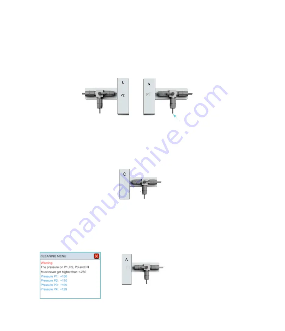
22. Attach a 5-10ml syringe at the extra P1 perfusion Inlet and push a small volume of buffer through
the 3-way valve to remove air in the valve. Then close the 3-way valve toward the P1 buffer flask as
shown below.
23. Now very gently with the syringe push buffer into the chamber (MONITOR the P1, P2 and P4 Pressure
on the Pressure Interface screen and DO NOT exceed 200mmHg in the CLEANING MENU). Push 1ml
into the chamber and P1 cannula to remove air. Close the 3-way valve toward the chamber as shown
below.
Attach a 5-10ml syringe with 5-10
ml running buffer


