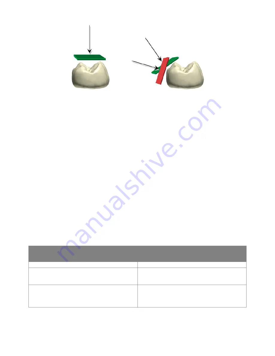
SCANNING PATH
The recommended scanning path consisting of 3 sweeps: occlusal, lingual and buccal to ensure good data
coverage of all surfaces.
The occlusal sweep always comes first as it has most of the 3D structure, which makes scanning
easier. The first sweep should be started at the first molar (if antagonist) or the preparation (to scan
gingiva before it collapses from the retraction). Allow the scanner to get a good ‘starting point’ by waiting
for 3-5 clicks before moving the scanner steadily and slowly 0-5mm above the teeth.
While scanning, the biggest challenge is to control the soft tissue, such as the tongue, lips and cheeks as
they may confuse the scanner if getting into its view and potentially slow down or even stop the scanning
process. Therefore, the easiest second sweep depends on the jaw:
•
The upper jaw has only the soft tissue on one side (buccal) therefore, the second sweep should be
buccal as this pushes the soft tissue away and creates a clear view for the scanner.
•
The lower jaw is more challenging because of the tongue. The cheek can easily be retracted using a
finger or a mirror. Therefore, the second sweep is lingual, pushing the tongue away.
•
The third sweep covers the opposite side of the second sweep. Again, try to avoid soft tissue. Because the
scanner has already been on the other side of the teeth during the sweep two, the system uses the
obtained data to avoid adding soft tissue to the scanned teeth.
The recommended scan paths are summarized.
General Principles:
Upper jaw
Lower jaw
1. Occlusion
1. Occlusion
2. Buccal - There is no soft tissue in the way.
2. Lingual - the tongue is the most moving soft
tissue (compared to the cheek). The cheek is
easily retracted.
3. Palatal - As the scanner has already been on
the other side of the teeth during the step two,
the system uses the obtained data to avoid
adding soft tissue to the scanned teeth.
3. Buccal - As the scanner has already been on
the other side of the teeth during the step two,
the system uses the obtained data to avoid
adding soft tissue to the scanned teeth.
If a scanned jaw has a preparation - start with the preparation, then follow the steps described above.
43


