Section 8- Protein A280
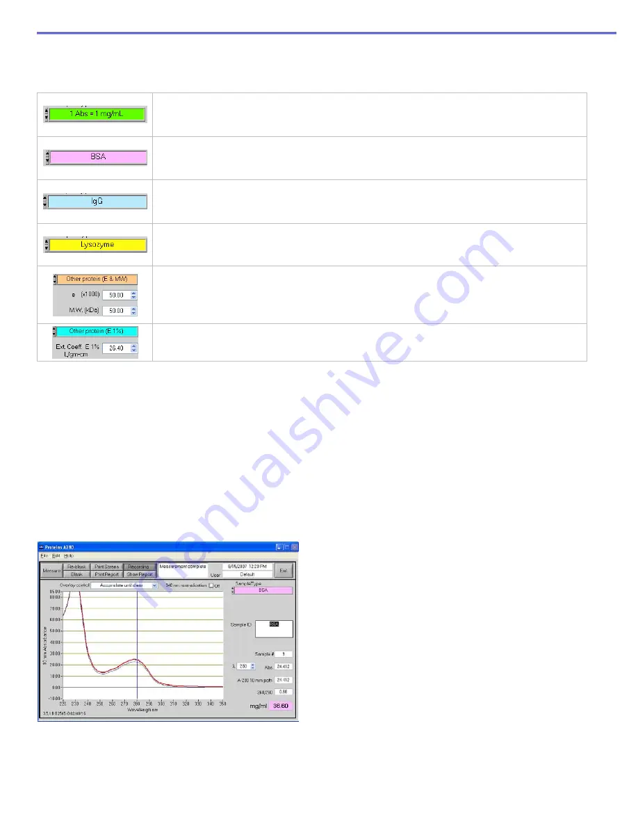
The sample type (color-keyed) can be selected by clicking on the preferred option or by scrolling through the selections
using the up or down arrow keys located to the left of the sample type box. A description of each sample type is given
below.
A general reference setting based on a 0.1% (1 mg/ml) protein solution producing an Absorbance at
280 nm of 1.0 A (where the pathlength is 10 mm or 1 cm).
Bovine Serum Albumin reference. Unknown (sample) protein concentrations are calculated using the
mass extinction coefficient of 6.7 at 280 nm for a 1% (10 mg/ml) BSA solution.
IgG reference. Unknown (sample) protein concentrations are calculated using the mass extinction
coefficient of 13.7 at 280 nm for a 1% (10 mg/ml) IgG solution.
Lysozyme reference. Unknown (sample) protein concentrations are calculated using the mass
extinction coefficient of 26.4 at 280 nm for a 1% (10 mg/ml) Lysozyme solution.
User-entered values for molar extinction coefficient (M
-1
cm
-1
) and molecular weight (MW) in kilo
Daltons for their respective protein reference. Maximum value for e is 999 X 1000 and maximum
value for M.W. is 9999 X 1000.
User-entered mass extinction coefficient (L gm
-1
cm
-1
) for a 10 mg/ml (1%) solution of the respective
reference protein.
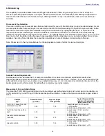
λ
and Abs:
current value of the user-selectable wavelength cursor and corresponding absorbance. The wavelength can
be set by dragging the cursor, using the up/down arrows or typing in the desired wavelength. Note: The user-selected
wavelength and absorbance are not utilized in any calculations.
A280 10-mm Path:
10 mm-equivalent absorbance at 280 nm for the protein sample measured
A260/280:
ratio of sample absorbance at 260 and 280 nm
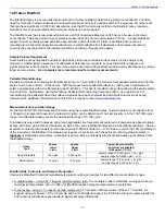
Spectrum Normalization
The baseline is automatically set to the absorbance value of the sample at 340 nm, which should be very nearly zero
absorbance. All spectra are referenced off of this zero. All data are now normalized and archived in the same format. The
user may elect to turn off the baseline normalization, which may result in the spectra being offset from the baseline.
Note: If the spectra baseline offset is significant, the calculated protein concentration may be higher than the true value.
Some samples may exhibit a greater baseline offset than the example above.
8-2


