background image

8

BUTTONS

PHACON

3D LIFE MODELING & SIMULATION

Buttons

 3

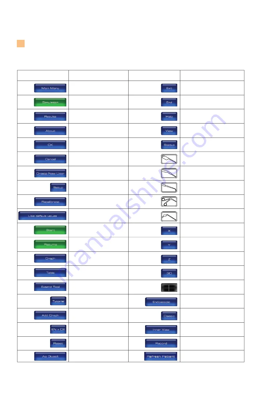
      Button                        Definition 

                   Button               

Definition

Login as guest.

Reload detected patient CT-data.

Access the MAIN MENU screen to view

major menu options.

Access the SIMULATION screen to begin

a training session.

Access the RESULTS screen to view and

compare the training session data.

Access the ABOUT dialog box to view

hardware and software information.

Accept inputs, changes or selections

and return to the previous screen.

Reject inputs, changes or selections and

return to the previous screen.

Access the CREATE NEW USER dialog

box to create a new user profile, user

name and password. 

Access the SETUP panel to perform

calibration of an instrument or patient.

Initiate the calibration of instruments or 

patient from the SETUP panel.

Select the factory default calibration set-

tings programmed for the instrument or 

Patient.

Begin a SIMULATION or training session

from the SETUP panel.

Continue a SIMULATION or training

session from the SETUP panel.

Access and view the training session

results in a graphical format.

Access and view the training session

results in a tabular format.

Virtual extension of the instrument. 

Open a tutorial.

Add a graph in the result screen.

Select or toggle the language for the 

software (German / English).

Reset the default settings for brightness, 

contrast and slice position in the 4 

windowpane in the simulation mode. 


Close and exit the program.

End the SIMULATION or training

session.

Access the HELP dialog box to view

instructions for use information.

Access the VIEW panel to select or

modify a screen view or perspective.

Access the STATUS panel to view the

number of injured risk structures during

a training session.

Select the POINTER for recalibration

and use. 

Select the angled drill for recalibration

and use. 

Select the ballprobe for recalibration

and use.

Select the blakesley for recalibration

and use.

Select the suction device for recalibration

and use.

Access the sagittal view of the skull CT

data as a full screen display.

Access the coronal view of the skull CT

data as a full screen display.

Access the transversal view of the skull

CT data as a full screen display.

Access the 3-D view of the Patient and skull 

as a full screen display.

Return to the default 4-windowpane

screen view.

View a navigated instrument in the 3-D

windowpane as if a virtual camera was

mounted on the tip of the instrument.

View a navigated instrument without the

virtual camera option.

Access the camera view onto the transparent 

cochlea.

Record a video of the camera view of the 

cochlea. 

Содержание SINUS TRAINER

Страница 1: ...USER MANUAL PHACON SINUS TRAINER...

Страница 2: ......

Страница 3: ...your trackers your tracking camera your patient your pointer your holder tray with skull your PHACON navigation software DISCOVER THE INTRODUCTION IN OUR PHACON TRAINER...

Страница 4: ...simulation 15 Select the axis view 15 Adjust the quality or perspective of the X Y and Z windowpane display 16 Adjust the perspective of the 3 D windowpane display 16 Change the view mode of the 3 D...

Страница 5: ...kull 23 Clean the skull holder with tray 23 Clean the trainer accessories 24 TROUBLESHOOTING 24 Troubleshooting 26 Error Messages 27 HOW TO PACK THE TRANSPORT CASE 28 PRODUCT WARRANTIES 29 MANUFACTURE...

Страница 6: ...camera F Sinus skull G Holder Tray with adjustable skull fixation H Ground cable 3x I Instrument tracker for ball probe J Pointer K Instrument tracker for suction M 3 x Instrument tracker for differe...

Страница 7: ...e image within the navigation camera s line of sight D Simulation Time Used to display the start time of the simulation the current time and the duration of the simulation session E Windowpanes 4 Used...

Страница 8: ...al Add a graph in the result screen Select or toggle the language for the software German English Reset the default settings for brightness contrast and slice position in the 4 windowpane in the simul...

Страница 9: ...new user click on the CREATE NEW USER button A CREATE NEW USER dialog box will appear Enter the required information into each field and then click OK 3 After a successful LOGIN an existing group can...

Страница 10: ...utton to perform instrument and patient calibration for optimal navigation results 6 See To Perform Setup for Navigation section 7 Click on the START button to begin simulation without performing cali...

Страница 11: ...rument click on icon Tool calibration RECALIBRATE Fig 5 Fig 5 ALWAYS make sure the round shapes of the pointer drill tracker or instrument tracker are aligned and visible to the navigation camera s li...

Страница 12: ...cedure 1 Click on the RECALIBRATE button to begin the calibration process Fig 6 2 Hold the tip of the pointer on the first calibration point of the Patient as indicated in the 3 D windowpane 3 After v...

Страница 13: ...egin the calibration process Fig 5 page 11 2 Make sure the desired tool is selected for calibration 3 Hold the tip of the tool in a fixed location on the Patient Make sure the tracking shapes are faci...

Страница 14: ...utton to access the VIEW panel options including AXIS VIEW CONTROL VIEW MODE and the OPACITYVALUE of an ANATOMICALSTRUCTURE Fig 8 NOTE The cross hairs visible within each windowpane represent the loca...

Страница 15: ...the mouse up and down To adjust the contrast click on the desired windowpane hold down the left mouse button and move the mouse left and right To adjust the zoom click on the desired windowpane hold d...

Страница 16: ...t key and left mouse button and move the mouse up and down or left and right The middle mouse button may also be used Fig 10 CHANGE THE VIEW MODE OF THE 3 D WINDOWPANE DISPLAY NOTE The ENDOSCOPIC view...

Страница 17: ...erformed to show the tool path during the simulation To virtually extend the tool in the 3 D windowpne click on the EXTEND TOOL button and move the slider to variable extend the virtual tool extension...

Страница 18: ...etter visualization within and behind the Patient 1 Click on the drop down list and select the desired anatomical structure Fig 12 2 Move the slider to change the opacity of the selected structure or...

Страница 19: ...to see which risk structure was injured and the time the injury occurred Fig 13 The start time current time and du ration of the training session is also displayed END THE SIMULATION OR TRAINING SESSI...

Страница 20: ...GRAPH or TABLE button to display the training session data in a graphical or tabular for mat respectively Fig 15 3 Click on the MEASUREMENT drop down list to select DURATION MINUTES or NUMBER OF INJU...

Страница 21: ...will allow access to the ANATOMICAL STRUCTURE drop down list Fig 16 4 Click on the ANATOMICAL STRUCTURE drop down list 5 Select the ALL option or a specific risk structure to view the associated numbe...

Страница 22: ...the GROUP drop down list and select a group 3 Click on the STATISTICAL PARAMETER drop down list and select a statistical parameter NOTE The STATISTICAL PARAMETER drop down list includes maximum minimu...

Страница 23: ...hin the skull using a dry brush and an external vacuum source Make sure the spillways are clear to allow water to low freely during the use of irrigation 5 Wipe all the surfaces of the skull including...

Страница 24: ...er is not detecting critical risk structures The ground cable with magnet is not installed on the instrument tracker or is not making contact with the manual instrument body Make sure the ground cable...

Страница 25: ...menu with a right click on your desktop and choose NVIDIA Control Panel 2 Select the Manage 3D settings in the left column of the NVIDIA Control Panel 3 In the Global Settings klick on High performan...

Страница 26: ...characters The entered group name already exists in the trainer Please enter a unique group name The entered group name is already used Enter a new group name into the trainer maximum of 150 characte...

Страница 27: ...and position the two camera arms into the recess next to the Tray the lenses facing down wards Fig 1 2 Cover the camera with the second layer of foam containing a recess for the tracker box Fig 2 3 In...

Страница 28: ...s if Products are used with accessories not manufactured by PHACON or if repairs are performed by any party other than authorized PHACON repair personnel Except as set forth above phacon makes no warr...

Страница 29: ...29...

Страница 30: ...30...

Страница 31: ...31...

Страница 32: ...PHACON GmbH Karl Heine Str 15 04229 Leipzig Germany Tel 49 0 3 41 47 83 97 32 Fax 49 0 3 41 47 83 91 74 www phacon de...

Отзывы:

Похожие инструкции для SINUS TRAINER