9
3.3 DNA Delivery Into Cells Using Electroporation (cont’d)
3.3.1 General Electroporation Discussion (cont’d)
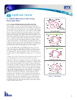
Research in the late 1980s and early 1990s showed that certain experimental conditions and parameters of
electrical pulses may be capable of causing many more molecules to move per unit time than simple diffusion.
There is also good evidence (Sukharev et al., 1992) that DNA movement is in the opposite direction of the arrow
in the sidebar.
An additional important consideration is when the voltage pulse is applied to the cells and medium that the
amount of current that flows is dependent on the conductivity of the material in which the cells are located.
Some material is quite conductive and severe heating will occur if the pulse duration is too long. Therefore long
duration fields will kill cells by destroying the membrane and heating.
The electric field in which the cells are located is produced by two system components. The first is the voltage
waveform generator and the second is the electrode which converts the voltage into the electric field.
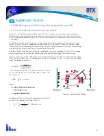
Neumann, Sowers and Jordan, 1998, pages 68-73 provides the equation that relates the transmembrane voltage
(TMV) to electric field intensity. As the charge accumulates at the membrane, which is a capacitance, the voltage
across the membrane increases.
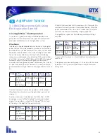
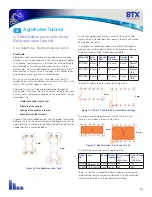
Figure 3-2: Transmembrane Voltage
voltage
charge
capacitance
=
As the voltage increases from its quiescent value of a few tenths
of a volt to more than 0.5 volts, pathways begin to form. The
TMV is given by:
TMV
3
⁄
2
-
E r |
cos
a
|
where:
E = electric field intensity in volts/cm
r = the cell radius in cm
a
= angle off the center line
To produce a TMV of 1 volt across the membrane of a cell with 7
µm radius, the required electric field intensity is:
2
⁄
3
=
=
950
volts / cm
1
7 *10
-4
E
-
AgilePulse
™
Tutorial
3


