24
23
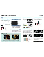
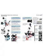
TIRFM (Total Internal Reflection Fluorescence
Microscopy) system with arc lamp source
Featuring the Olympus-developed total internal reflection
illumination system and slit mechanism to provide evanescent wave
illumination from an arc lamp source. High signal to noise
fluorescence observations with extremely thin optical sectioning can
now be easily performed at the specimen-coverslip interface.
The arc lamp is focused on an off-center slit using a wedge prism
and focused on the outer edge of the back focal plane of the
objective, thus causing the excitation light to exit the objective
beyond the critical angle resulting in Total Internal Reflection.
The wedge prism and slit can be easily removed from the light path
via a slider for wide field fluorescence observation. Through the use
of filters, this system
enables a wider choice of
excitation colors than
current laser base
system.
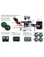
Conventional fluorescence observation
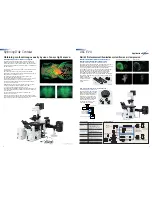
Microtubule of an NG108-15 cell labeled with Alexa488 through indirect fluorescence antibody test
TIRFM observation
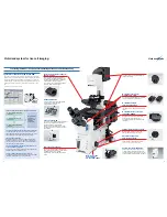
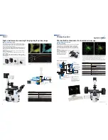
High-precision fluorescence turret /IX2-RFACEVA
Turret includes three, highly precise,
empty fluorescence filter cubes that
permit dichromatic mirror switching
while maintaining excitation light
position on the back focal plane of
the objective. This system makes
multi-color observations easy and
alleviates the additional adjustment
of the excitation source when
switching mirror units. Up to six
mirror units can be installed.
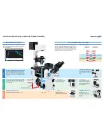
Off-center slit slider
Excitation filter slider
Fluorescence lamp housing
L shape fluorescence
illuminator
Exclusive
objective
High precisiton
fluorescence turret
Wedge prism slider
LIFE TIME
BURNER ON
LIFE TIME
BURNER ON
U-LH75XEAPO
75 W xenon apo
lamp housing
U-RFL-T
Power supply unit
for mercury lamp
U-RX-T
Power supply unit
for xenon lamp
U-LH100HG
100 W mercury
lamp housing
U-LH100HGAPO
100 W mercury apo
lamp housing
Mirror unit
IX2-RFAL
L shape fluorescence
illuminator
IX2-RFAC
Fluorescence turret
Camera
adapter
High
sensitive
camera
IX71
Research
inverted
system
microscope
APON60XOTIRF
objective
Exclusive
vacant mirror unit
High-precision
fluorescence turret
Excitation filter slider
Motorized filter wheel
Wedge prism slider
Off-center slit slider
IX2-RFACEVA
IX2-ARCEVA
Kaede-Crk II protein expressed in a HeLa cell
IX2-ARCEVA
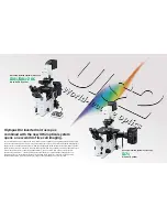
World’s first evanescent illumination system from an arc lamp source.
ARC EVA
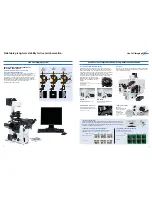
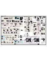
SYSTEM DIAGRAM
Main specifications
Microscope
Research inverted system
microscope IX71
Fluorescence
Arc illumination total internal reflection
illuminator
fluorescence unit IX2-ARCEVA
(Slit slider, wedge prism slider and
excitation filter slider)
L-shape fluorescence illuminator
IX2-RFAL
Mirror unit cassettes
High-precision fluorescence turret
(choose from either
IX2-RFACEVA
fluorescence turret)
(with centering mechanism and
3 vacant mirror units)
Fluorescence turret
IX2-RFAC
Lamp light source
100 W mercury lamp,
75 W Xenon lamp
Objective
APON60XOTIRF
N.A. 1.49. W.D. 0.1 mm
Used with normal cover glass and
immersion oil
Stage
Left short handle stage IX-SVL2
Total internal reflection
11
illumination F.N.
Observation
Recommend high sensitive camera
Application System
Disk Scanning Confocal Microscope System
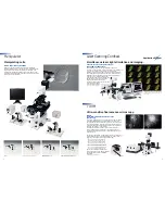
The Olympus Disk Scanning Unit (DSU) offers confocal images
using a white light, arc excitation source and CCD camera. The
heart of the system is a unique slit disk pattern, that offers excellent
light throughput and thinness of optical Sectioning. Compatible with
any IX71 and IX81.
• Compliance with various fluorochromes with different spectral
characteristics.
Since an arc light source is used, the unit can meet different
fluorochrome requirements across a wide wavelength spectrum by
simply switching a standard mirror unit.
• Minimize excitation light damage to the specimen and
maximize emission light throughput.
The excitation light volume is reduced to around 5% as a result of
passing through the disk. So, there is almost no fading of
fluorescence emission from the surface of the focused sample.
• Construction of 3D images.
Brilliant 3D image can be easily captured with excellent optical
sectioning with high precision motorized Z axis of IX81.
• Low and high magnification objective support.
Five DSU disks are available of varying slit spacing and width for the
wide variety of the objectives, included oil or water immersion high
N.A. objectives.
• Easy switching between confocal and reflected light
fluorescence observation .
IN/OUT of the confocal disk to or from the light path can be done
by a hand switch or via software, so it is easy to switch observation
methods between DSU and reflected light fluorescence.
Spinning Disk Confocal
Zebrafish 3-day embryo, ventral view, projection of 62 serial optical sections
Adult brain of
Drosophila
, reflected light fluorescence image (left) and DSU image (right)
Obtaining confocal images easily by use of an arc light source.
* Not available in some areas
* Not available in some areas

