22
Chapter 3 Cleaning, Disinfection and Sterilization Procedures
EVIS EXERA GIF/CF/PCF TYPE 160 Series REPROCESSING MANUAL
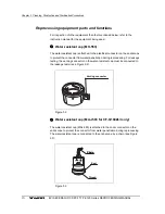
Inspection of the channel plug
Confirm that the cylinder plug and biopsy valve cap are free from cracks,
scratches, flaws and debris (see Figure 3.5).
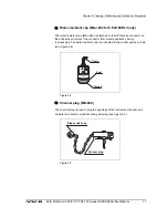
Inspection of the injection tube
1.
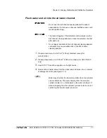
Confirm that all components of the injection tube are free from cracks,
scratches, flaws and debris (see Figure 3.6).
2.
Confirm that the filter mesh is in the suction port.
3.
Attach the 30 cm
3
(30 ml) syringe to the air/water channel port. With the
filter mesh immersed in rinsing water, withdraw the syringe plunger and
confirm that rinsing water is drawn into the syringe. Depress the plunger and
confirm that rinsing water is emitted from the air pipe port. Confirm that
water is not emitted from the suction port.
4.
Attach the 30 cm
3
(30 ml) syringe to the suction channel port. With the filter
mesh immersed in rinsing water, withdraw the syringe plunger and confirm
that rinsing water is drawn into the syringe. Depress the plunger and confirm
that rinsing water is emitted from the distal end of the suction channel tube.
Confirm that water is not emitted from the suction port.
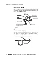
Inspection of the auxiliary water tube (for endoscopes with
auxiliary water feeding only)
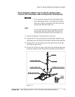
Check for cracks, scratches, flaws, debris and other damage (see Figure 3.7).
Inspection of the channel cleaning brush
1.
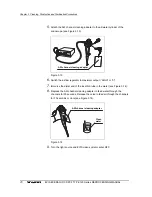
Confirm that the brush section and the metal tip at the distal end are
securely in place. Check for loose or missing bristles (see Figure 3.8).
2.
Check for bends, scratches and other damage to the shaft.
3.
Check for debris on the shaft and/or in the bristles of the brush.


