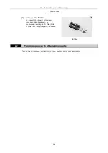


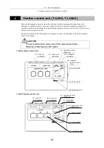

III
Detailed Sequence of Microscopy
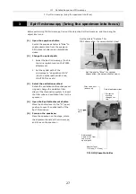
3 Epi-fl microscopy (bring the specimen into focus)
28
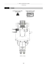
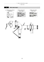
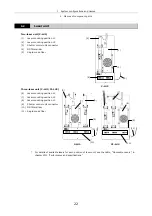
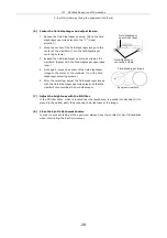
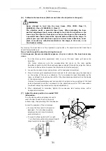
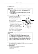
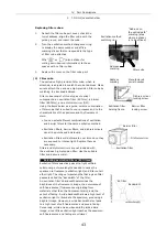
(6) Center the field diaphragm and adjust its size.
1 Reduce the field diaphragm opening. (Move the field
diaphragm open/close lever to the “C” (close)
direction.)
2 Move the center of the field diaphragm image to the
center of the viewfield. (Turn the field diaphragm
centering screws.)
3 Expand the field diaphragm opening as wide as the
viewfield. (Adjust with the filed diaphragm open/close
lever.)
4 Once again, move the center of the field diaphragm
image to the center of the viewfield. (Turn the field
diaphragm centering screws.)
5 After the centering, adjust the field diaphragm image
with the field diaphragm open/close lever so that the
viewfield circumscribes the field diaphragm.
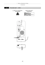
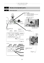
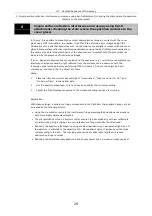
(7) Adjust the brightness with the ND filter.
If the ND filter slider, which is located near the lamphouse, is pushed into backward, it is
placed in the optical path, thus reducing the brightness of the image.
(8) Close the Epi-fl attachment shutter.
In order to prevent fading of the specimen, always close the shutter for Epi-fl illumination
when interrupting the Epi-fl microscopy.
Field diaphragm
centering screws
Field diaphragm
open/close lever
Eyepiece viewfield
Field diaphragm image
Содержание TIRF2
Страница 1: ...TIRF2 SYSTEM FOR TE2000 INSTRUCTIONS M339E 04 12 NF 2 ...
Страница 2: ......