OPERATION
Axio Lab.A1
Lighting and contrasting method in transmitted light
Carl Zeiss
04/2013 430037-7144-001
93
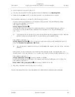
(3) Setting the microscope
x
Set the microscope as for the transmitted light brightfield (see Section 4.1.1), taking care to ensure the
correct interpupillary distance in the binocular tube (see Section 3.5.5).
x
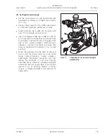
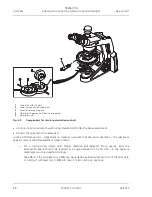
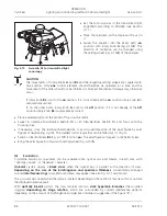
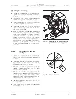
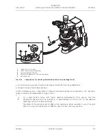
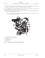
Center rotary stage Pol (Fig. 4-5/
1
) and objectives (see Sections 3.1.8.5 and 3.1.8.6).
x
Swivel polarizer (Fig. 4-5/
3
) into the light path and, if it is rotatable, position it at 0°.
x
Swivel the analyzer into the beam path and bring it into a crossed position using the setting wheel
(Fig. 4-5/
2
). The field of view will appear dark due to the crossed polarizers.
x
Set the alignment specimen Pol on the microscope stage and turn to the dark position of the
alignment specimen.
x
Swivel out the analyzer and align the crossline reticle with the cracks in the specimen.
x
Subsequently swivel the analyzer back in and remove the alignment specimen. The pass directions of the
polarizer and analyzer will now be parallel to the crossline reticle (polarizer EW, analyzer NS).
An adjustment of the crossline reticle is not necessary when working with the intermediate
plate and the binocular photo tube Pol (425520-9100-000).
x
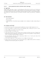
Rotate the rotary stage Pol with the specimen, e.g. a synthetic fiber, until the specimen appears as
dark as possible. In this position, the fiber extends parallel to one of the two directions of the crossline
reticle.
Do not change the interpupillary distance on the binocular tube, as the the angle of the
crossline reticle to the fiber will be changed.
x
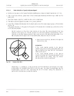
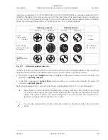
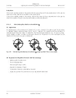
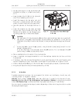
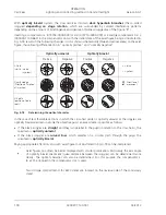
Now turn the stage on by 45° so that the longitudinal axis of the fiber is oriented NE-SW (Fig. 4-15).
The specimen will display the greatest brightness here (diagonal position). In this position the
specimen may have any color.
x
Inserting compensator
O
(473704-0000-000).
Like the specimen, the compensator
O
is a birefringent object, albeit with a defined path difference of
550 nm and the principal direction of oscillation n
J
definitely oriented in a NE-SW direction.
By moving compensator
O
into the light path, the specimen changes its color. The type of color change
depends on the orientation of the specimen (NE-SW or NW-SE).
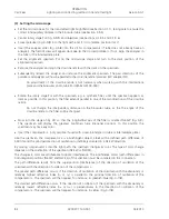
The changes in color are attributable to optical interference. The interference colors (path differences) in
both diagonal positions (NE-SW and NW-SE) of the specimen must be compared in this connection.
The path difference results from the superposition (interference) of the direction of oscillation of the
specimen with the direction of oscillation of the compensator
O
.
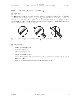
The greater path difference occurs, if the direction of oscillation of the specimen with the absolutely or
relatively highest refractive index (n
J
or n
J
’
) is parallel to the principal direction of oscillation of the
compensator
O
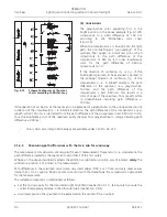
. The specimen will then appear, for instance, in greenish-blue (Fig. 4-14/
2
).
The smallest path difference occurs, if the direction of oscillation of the specimen with the absolutely or
relatively lowest refractive index (n
D
or n
D
’
) is perpendicular to the direction of oscillation of the
compensator
O
. The specimen will then appear, for instance, in yellow (Fig. 4-14/
3
).

