35
IX71/IX51
}Before proceeding to the following, open the aperture iris diaphragm
because flare would be observed at the center when it is stopped down.
1. Engage the phase contrast objective in the light path and bring the
specimen into focus.
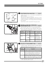
2. When the U-BI90CT binocular observation tube is used, rotate the CT
turret @ to position “CT”. When the observation tube in use is other than
the U-BI90CT, remove an eyepiece and attach the U-CT30 centering
telescope in place. (Fig. 51)
3. Engage the ring slit of the condenser matching the phase contrast
objective in the light path.
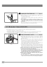
4. Rotate the focus ring ² (or the knurled section when the U-CT30 is used)
to focus on the ring slit ³ and the phase plate | of the objective.
(Figs. 51 & 52)
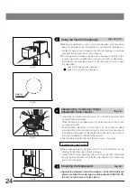
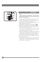
5. Using the optical element centering knobs ƒ, turn the phase contrast
ring slit centering screws (in positions marked ) so that the ring slit
image overlaps with the phase plate of the objective.
}A ghost of the ring slit image may be observed. In this case, overlap the
brightest image with the phase plate.
}If a thick specimen is moved, the ring slit image may be deviated from the
phase plate and the contrast may be deteriorated. In this case, re-adjust the
centering by repeating steps 1 to 5 above.
6. After completing centering, rotate the CT turret to return the turret to
position “0”. If the centering telescope is in use, replace it with the
eyepiece.
}If the vessel is not completely flat, it may become necessary to adjust the
centering again to obtain the optimum contrast.
Repeat centering by beginning with the lowest-power objective and
increasing the objective power in order.
7. Adjust the field iris diaphragm so that its image circumscribes the field of
view and observe the phase contrast.
}Engaging the green filter in the light path will improve the contrast.
Fig. 51
@
²
Fig. 52
³
|
Fig. 53
ƒ
3
Centering the Phase Contrast Ring Slit
(Figs. 51 to 53)

