INTERSON ViewBladder 10 User Manual Download Page 13

01-9932-00 ViewBladder 10 User Guide Revision 2                                                                                             12 
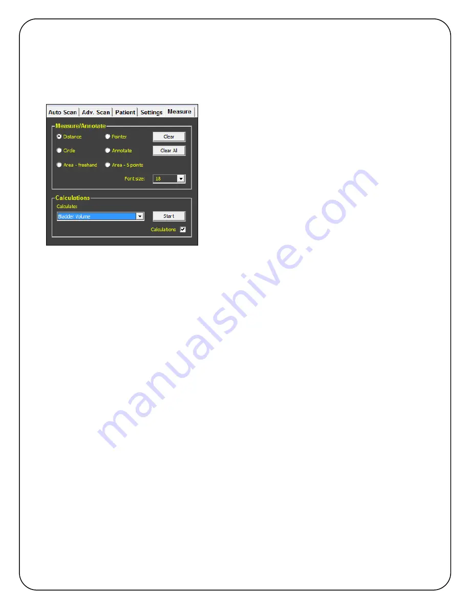
 

6.5.  Measure Tab 


The

 Measure 

tab is used to add measurements and annotations to an image, as well as perform  

Bladder Volume and Bladder Volume 3D calculations.  Contact Interson for additional calculations 
that can be added to your system.  Prostate Volume, Crown Rump, Gestational Sac, Femur 
Length, Head Circumference, Abdominal Circumference, and Bi-Parietal Diameter are available. 


È


There are four types of measurements available. 
 

Distance

 is invoked by placing your finger on a starting 

point and dragging your finger to the end point and then 
lifting your finger. 


Similarly, a perfect 

Circle

 can be drawn.  Place your 

finger at one edge of the circle and drag your finger to 
the other edge of the circle. 


To draw a random shape use 

Area-freehand


To draw a smooth shape, use 

Area - 5 points

 and 

select five points on the image.  ViewBladder 10 will 
smoothly connect the five points. 


Annotate

 and 

Pointer

 are used to label items on the image. 


Font Size

 can be changed to suit your preference. 


Clear

 removes the most recent measurement or annotation one at a time. 


Clear All

 removes all calculations, measurements, and annotations. 


To enable calculations, select the white box in the 

Calculations

 window.  There are two 

calculations available in ViewBladder 10.  Use the 

Calculate

 pull down to select either Bladder 

Volume or Bladder Volume 3D.  Bladder Volume uses the height and width of a transverse 
bladder scan to estimate bladder volume.  Bladder Volume 3D adds a thickness measurement 
from a mid-sagittal scan to the height and width measurements from the transverse scan.  
Bladder Volume 3D use the industry-standard Height x Width x Thickness x 0.7 to estimate the 
bladder volume. 
 
Pressing 

Start

 begins the calculation procedure and provides text prompts underneath the 

calculations window. 

Summary of Contents for ViewBladder 10

Page 1: ...terson is a trademark of Interson Corporation ViewBladder 10 is a trademark of Interson Corporation SeeMore USB Probe is a trademark of Interson Corporation Equipment Manufacturer M EC Representative...

Page 2: ...aining 5 4 GENERAL 6 5 VIEWBLADDER 10 DISPLAY 6 6 VIEWBLADDER 10 USER CONTROLS 7 6 1 Auto Scan Tab 7 6 3 Patient Tab 10 6 4 Settings Tab 11 6 5 Measure Tab 12 7 SAVING VIEWING AND PRINTING IMAGES REPO...

Page 3: ...sor 1 5 GHz x Minimum RAM 2 GB RAM x USB 2 0 port 3 Warnings Safety Information 3 1 Meaning of Signal Words In this User s Manual the signal words Warning and Caution are used regarding safety and imp...

Page 4: ...stem must have a general knowledge of the use of ultrasound imaging devices and imaging protocols Connect the probe to the USB port prior to turning on the tablet Do not use in the presence of flammab...

Page 5: ...2 Probe Disinfecting A 10 6 reduction in pathogens should be reached following the disinfecting procedures in this manual and using the following recommended solutions The following disinfectants are...

Page 6: ...used however never apply cleaner directly to the tablet surface Dampen the microfiber towel with the cleaner Do not saturate the towel 3 7 Surface Disinfection The tablet surface can be disinfected u...

Page 7: ...sure the width The calculated volume is now displayed on the left of the screen Directly below the Imaging Window is the Status Window If you have started a patient exam the left hand column shows all...

Page 8: ...page report The following steps will guide you through acquiring a Bladder Volume using three distance measurements First you will acquire a transverse image of the bladder to measure the height and w...

Page 9: ...ve the optimal bladder image 8 You should see a black ultrasound shadow on the right edge of the scan This is the patient s pubic bone 9 Measure the thickness of the bladder by placing your finger on...

Page 10: ...nd parameters A specific preset contains Intensity Contrast Near Gain Mid Gain Far Gain Frequency Depth and Power The list of available presets changes based on the probe that is selected To save a ne...

Page 11: ...hecked patients are displayed in the order they were entered Patient Exams are initiated and completed from the Auto Scan tab After an exam is completed Print will print a single page report for the s...

Page 12: ...ce line can be displayed if an endocavity probe is the specified probe Cine Frames allows the user to specify the number of frames that are buffered for replay 32 64 128 or 256 may be selected As the...

Page 13: ...e use Area 5 points and select five points on the image ViewBladder 10 will smoothly connect the five points Annotate and Pointer are used to label items on the image Font Size can be changed to suit...

Page 14: ...d graphics program to open and print any previously saved jpeg image Navigate to the Patient Folder using the shortcut on the desktop and select the jpg file that you would like to print You can also...

Page 15: ...Group 1 Class a Under Limit CISPR 11 Measure at 5 meters Electrostatic Discharge EN60601 1 2 2kV 4kV 8kV contact discharge 2kV 4kV 8kV air discharge 36 202 1 j EN61000 4 2 Apply to all accessible com...

Page 16: ...0 to 80 no condensation x Atmospheric pressure 700 hPa to 1060 hPa 10 Transportation Never carry the probe by the cable The cable could disconnect from the probe allowing it to drop and possibly damag...

Page 17: ...Periodic testing and maintenance of the Interson USB ultrasound probe is NOT required YWARNING Do not use cables or accessories other than those provided with the Interson USB Ultrasound Probe as thi...

Page 18: ...to one of the computer s USB 2 0 ports 2 Navigate to the location that contains the file ViewBladder 10 Setup exe 3 Click on the icon ViewBladder 10 Setup exe 4 Follow the on screen instructions clic...

Page 19: ...ransducers High Bandwidth single element 3 5 MHz 5 MHz 7 5 MHz 12 MHz 15 MHz and 24 MHz Depth Selections 3 5 6 10 15 and 20 cm depths Measurements Distance area volume measurements Signal Processing x...

Page 20: ...ystem Architecture System Specification Requirements for PC Tablet or Laptop Processor 1 5 GHz or higher Memory 2 Gigabyte or more Video Chipset Intel 815EM or higher performance equivalent NVIDIA rec...

Page 21: ...atomy Focal Point 2 0 cm Max depth 10 0 cm Patient contact area 20 0 mm Displayed depth 5 cm 10 cm USB PROBE MV 12 0 MHz Human Vascular Phlebotomy Focal Point 0 5 cm Max depth 3 0 cm Patient contact a...

Page 22: ...3 NA NA Spatial peak temporal average intensity mW cm2 13 5 13 5 NA NA 13 5 NA NA Attenuated spatial peak temporal average intensity mW cm2 3 98 3 98 NA NA 3 98 NA NA Peak rarefactional acoustic pres...

Page 23: ...mporal average intensity mW cm2 18 7 18 7 NA NA 18 7 NA NA Attenuated spatial peak temporal average intensity mW cm2 6 92 6 92 NA NA 6 92 NA NA Peak rarefactional acoustic pressure MPa 2 22 2 22 NA NA...

Page 24: ...l average intensity mW cm2 62 2 62 2 NA NA 62 2 NA NA Attenuated spatial peak temporal average intensity mW cm2 40 3 40 3 NA NA 40 3 NA NA Peak rarefactional acoustic pressure MPa 3 10 3 10 NA NA 3 10...

Page 25: ...l average intensity mW cm2 55 4 55 4 NA NA 55 4 NA NA Attenuated spatial peak temporal average intensity mW cm2 36 2 36 2 NA NA 36 2 NA NA Peak rarefactional acoustic pressure MPa 2 80 2 80 NA NA 2 80...

Page 26: ...oral average intensity mW cm2 35 7 35 7 NA NA 35 7 NA NA Attenuated spatial peak temporal average intensity mW cm2 18 9 18 9 NA NA 18 9 NA NA Peak rarefactional acoustic pressure MPa 3 16 3 16 NA NA 3...

Page 27: ...al average intensity mW cm2 3 64 3 64 NA NA 3 64 NA NA Attenuated spatial peak temporal average intensity mW cm2 3 49 3 49 NA NA 3 49 NA NA Peak rarefactional acoustic pressure MPa 1 45 1 45 NA NA 1 4...

Page 28: ...Intra Vascular Peripheral Vascular z z z Laparoscopic Muscular Skeletal Conventional z z Muscular Skeletal Superficial z z Others Specify 1 USB Transducer GP 3 5 MHz AB 3 5 MHz This device is a hand h...

Page 29: ...ent mechanical sector probe intended for transcutaneous use with the INTERSON USB ULTRASOUND PROBE SYSTEM The nominal operating frequency is 12 MHz In B mode the transducer operates over a 29 mm area...

Page 30: ...someone stole my laptop computer or storage device What would I do if my computer hard drive crashed Ultimately it is the user s responsibility to assure the security of their data Several strategies...

Page 31: ...terial or workmanship not provided or furnished by the Company ii the Product resulting from abnormal use of the Product or use in any manner other than as specified in the Product s operating manual...

Page 32: ...0 days of ownership To register your system for an extended warranty copy this page fill out your information and send to Interson with payment Customer _______________________________________________...

Reviews: