62
Appendix
A.1
X-ray Exposure Guide
The required X-ray dose for the best image is dependent on the following:
-
X-ray source (tube assembly, manufacturer, AC/DC, etc.)
-
Distance between beam focus and sensor
-
Tooth (object) to be X-rayed
-
Bone density and age of patient
-
Miscellaneous circumstances, etc.
The X-ray dose influences image quality. Based on fundamental laws of physics, an insufficient
dose generally means higher image noise, which leads to poor detail discrimination. On the
other hand, an excessively high dose can cause the sensor to be overexposed. This is also
perceptible by a decrease in detail discrimination, specifically in darker areas.
The effect of image processing reduces the difference between image qualities of different
doses. Users can adjust brightness and contrast in the option menu.
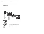
The recommended exposure dose is from 300μGy to 600μGy when measuring without an
object. Exposure time corresponding to the dose may vary depending on the X-ray equipment
used. Recommended exposure times according to positions are as shown on the Exposure
Time Table.
The X-ray dose is maintained through tube voltage (kVp) and current (mA), as well as exposure
time according to the signal level.
Since the exposure time depends on the diagnostic problem as well as the
clinical situation, the selection of an adjustment is the responsibility of the
treating physician.
Image degradations caused by overexposure of the sensor cannot be
compensated, but an insufficient dose can be partially compensated
through image processing.
https://stomshop.pro


