


•
Patients with a reduced ejection fraction should be on stable AHA/ACC guidelines
based medical therapy prior to implant.
•
The PA Sensor is a permanent implant. The sensor does not have any batteries that
require replacement or any components that will wear or fail over time. Removal after
implantation is not recommended. No circumstances where the sensor needs to be
removed have been identified and no sensor has been removed during the clinical
trial of the device. The sensor should be retrieved by using standard intra-vascular
and surgical procedures as would be used for other vascular implants if required.
•
If there is evidence of a change in device performance, contact Technical Support for
additional information.
•
PA Sensor function is unaffected after temporary exposure up to 2 Atmospheres
Absolute (ATA) pressure. Follow the contact process under Technical Support for
additional information if the patient will have hyperbaric chamber exposure or is
planning to scuba dive.
•
Pacemakers, ICD’s, and Ventricular Assist Devices (VAD) can work in conjunction
with the PA Sensor and will not affect the performance of the system. Several medical
procedures can also be performed while the sensor is implanted if precaution is taken
to avoid direct contact with the sensor. These procedures include radiofrequency
ablation, ionizing radiation, and diagnostic ultrasound. The effects of therapeutic
ultrasound have not been determined. If therapeutic ultrasound is required, avoid
contact with the sensor.
MRI Information
Non-clinical testing demonstrated that the sensor is MR Conditional. A patient with this
device can be scanned safely, immediately after implantation under the following
conditions:
o
Static magnetic field of 1.5 or 3.0 Tesla
o
Maximum spatial gradient magnetic field of 720-Gauss/cm (7200-mT/m)
or less
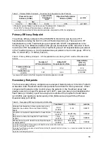
In non-clinical testing, the CardioMEMS PA Sensor produced the temperatures in the
table below during MRI performed for 15 minutes of scanning (per pulse sequence) in the
1.5-Tesla/64-MHz
and 3-Telsa/128-MHz
MR systems. These temperature changes will
not pose a hazard to the patient under the conditions indicated.
Table 2. MRI Related Heating
1.5-Tesla
3-Tesla
MR system reported whole body averaged SAR
2.9-W /kg
2.9-W /kg
Calorimetry measured values, whole body averaged SAR
2.1-W /kg
2.7-W /kg
Highest temperature change
1.9°C
2.3°C
MR image quality may be compromised if the area of interest is in the same area or
relatively close to the position of the sensor. Selecting optimal MR imaging parameters to
compensate for the presence of the sensor may be necessary. The maximum artifact size
(as seen on the gradient echo pulse sequence) extends approximately 5 mm relative to
the size and shape of the sensor.
1
Magnetom, Siemens Medical Solutions, Malvern, PA. Software Numaris/4, Version Syngo MR
2002B DHHS Active-shielded, horizontal field scanner
2
Excite, HDx, Software 14X.M5, General Electric Healthcare, Milwaukee, WI
7


