Collect data
Collect data
Connect the sensor to SPARKvue or Capstone to collect data.
SP
SPARK
ARKvue
vue
1. Turn on the sensor.
2. Open SPARKvue then click Sensor Data.
3. Select the that matches its device ID.
4. Under Quick Start Experiments, select EKG and Heart Rate
EKG and Heart Rate.
5. Click Start
to begin data collection.
P
PASCO Capst
ASCO Capstone
one
1. Turn on the sensor.
2. Open Capstone then click Hardware Setup
.
3. Select the that matches its device ID. Click Hardware Setup
to
close the panel.
4. Select the EKG and Heart Rate
EKG and Heart Rate Quick Start Experiment.
5. Click Record
to begin data collection.
Analyzing the electr
Analyzing the electrocar
ocardiogr
diogram
am
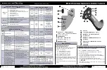
Figure 2. Example electrocardiogram.
The voltage versus time graph displays an electrocardiogram
(Figure
Figure 22). One part of the electrocardiogram is a trace indicating no
detectable electrical activity (flat line). This line is called the isoelectric
line. Deviation from the isoelectric line indicates electrical activity of
the heart muscles. The sensor’s voltage measurement represents this
deviation.
The first deviation from the isoelectric line is an upward pulse
followed by a return to the isoelectric line. This is called the P wave
P wave.
This wave is caused by the depolarization of the atria and is
associated with the contraction of the atria.
After a return to the isoelectric line, there is a short delay while the
heart’s atrioventricular (AV) node depolarizes and sends a signal along
the atrioventricular bundle of conducting fibers to the Purkinje fibers.
The Purkinje fibers bring depolarization to all parts of the ventricles
almost simultaneously.
After the AV node depolarizes there is a downward pulse called the Q
Q
wave
wave. Shortly after the Q wave there is a rapid upswing of the line
called the R wave
R wave. This is followed by a strong downswing of the line
called the S wave
S wave and then a return to the isoelectric line. These three
waves together are called the QRS complex
QRS complex. This complex is caused
by the depolarization of the ventricles and is associated the with the
contraction of the ventricles.
After a short period the sodium and calcium ions that have been
involved in the contraction migrate back to their original location in a
process that involves potassium ions and the sodium-potassium
pump. The movement of these ions generates an upward wave that
then returns to the isoelectric line. This upward pulse is called the TT
wave
wave and indicates repolarization of the ventricles.
The sequence from P wave to T wave represents one heart cycle. The
number of such cycles in a minute is called the heart rate and is
typically 70-80 cycles (or beats) per minute at rest.
Wireless EKG Sensor | 3