


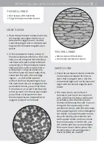
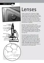
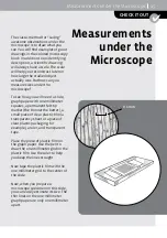

Eyepiece lens
Eye
Light source
Actual plane
of intermediate image
Intermediate
lens
Objective lens
Slide
Stage
Diaphragm disk
Apparent
image plane
0.0025 mm in reality
1 cm under the
microscope
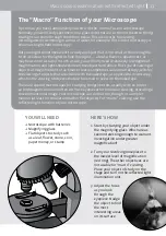
Drops of water and curved pieces of glass
have something in common: They magnify
objects when you look through them. Curved
pieces of glass are also known as lenses. Since
the 19th century, when Ernst Abbe began
improving the magnification power of lenses,
not just by trial and error, but also through
mathematical calculations, microscopes have
become better and better.
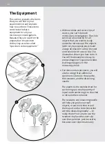
But what exactly does “better” mean?
Basically, it means getting a clearer and larger
image through more skillfully shaped lenses
and through the combination of various
lenses. In your microscope, you will always
be seeing the image through two lenses. One
lens is located in the eyepiece, the other in
the objective.
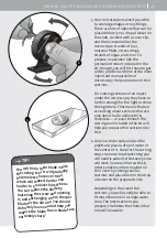
Each objective has a different lens, with
each lens magnifying the slide specimen
more strongly than the last. You can see
the magnification written on the revolving
nosepiece. Under the microscope’s highest
magnification, objects will appear 400 times
larger. That means that two cells that seem
to be 1 centimeter apart from each other
under the microscope will actually be just 25
thousandths of a millimeter apart.
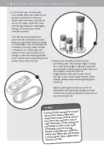
CHECK IT OUT
10
Lenses


