background image

2

1

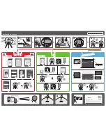
Rod is parallel 
in all cases.

Pins include 
tibial slope.

With the knee flexed at 90 degrees, place the tibial resection 
guide with uprod assembly onto the proximal anterior 
medial aspect of the tibia and both plateaus. Avoid using 
excessive force to seat the guide. Apply most of the force 
anterior to posterior while holding the guide as described. 

To assist in the medial/lateral positioning of the tibial pin 
guide, refer to the last page of the Patient Proposal which 
contains a top view of the patient’s tibial surface. It is 
recommended to visualize the red line shown in the 
Patient Proposal to the patient’s bone and to check 
alignment with the raised line on the lateral aspect of the 
tibial pin guide (Figure 4).

The planned Varus/Valgus (V/V) alignment can be confirmed 
by verifying the alignment of the rod to the patient’s tibial 
crest and center of the ankle (Figure 5). The rod is designed 
to be parallel to the mechanical axis of the tibia regardless of 
the planned tibial slope, when viewed laterally.

Note: 

The position of the line in the Patient Proposal 

is intended to reference the medial one-third of the 
tibial tubercle and not the middle of the tibial crest 
(Figure 4).

Note: 

It is recommended to clear extraneous tissue 

along the anterior medial aspect of the tibia. Soft 
tissue impingement can impact the fit of the guide 
and overall alignment or slope. Visualization in 
assessing proper fit observed from a sagittal or side 
view is helpful. 

Note: 

To position the guide, apply most of the 

pressure to the anterior aspect and the remaining 
pressure to the proximal aspect of the guide. This 
will help assure proper seating of the guide at the 
appropriate resection level. The correct position is 
found when there is minimal or no toggling/rocking 
of the tibial pin guide.

Once the tibia pin guide and uprod assembly is in the 
desired position, hold it in place, and secure it to the bone 
by drilling two (recommended P/N 9505-02-302) pins, first 
through the lateral and then the medial, drill guide pin 
holes (Figure 6).

Figure 4

Figure 5

Figure 6

0 degree block 
should be used as 
slope has been 
planned in the pin 
placement.

Surgical Technique  TRUMATCH Personalized Solutions Pin Guides  DePuy Synthes Joint Reconstruction    5

Содержание Depuy Synthes Trumatch

Страница 1: ...Pin Guide System SURGICAL TECHNIQUE TRUMATCH PERSONALIZED SOLUTIONS with the SIGMA High Performance Instruments...

Страница 2: ...operation following the use of the TRUMATCH Solutions Femoral and Tibial Pin Guides It is strongly recommended that the surgeon carefully review the TRUMATCH Solutions Patient Proposal prior to proce...

Страница 3: ...ition the distal femoral cutting block with anterior reference guide Step 5 Use of angel wing to verify distal resection level Tibial Preparation SIGMA Total Knee System steps shown Step 1 Insert dril...

Страница 4: ...The TRUMATCH Solutions Drill Guides P N 2004 20 925 are reusable after sterilization A minimum of four 4 drill guides should be on hand for a case They are shipped separately from the TRUMATCH Solutio...

Страница 5: ...ote The position of the line in the Patient Proposal is intended to reference the medial one third of the tibial tubercle and not the middle of the tibial crest Figure 4 Note It is recommended to clea...

Страница 6: ...confirm the cut orientation with the angel wing If necessary the block may be shifted 2 mm proximally or distally by selecting the appropriate offset holes adjacent to the 0 mm hole Perform the proxi...

Страница 7: ...and position the feet of the guide over the distal femoral condyles Figure 11A Avoid using excessive force to seat the guide Care should be taken to avoid squeezing the block and causing the legs to...

Страница 8: ...guide by flexing the guide from posterior to anterior Note The TRUMATCH Solutions Drill Guides P N 2004 20 925 are reusable after sterilization A minimum of four 4 drill guides should be on hand for...

Страница 9: ...the distal femoral resection using a 1 19 mm whale tail thick saw blade Figure 14B Remove the HP Distal Femoral cutting block and confirm the bone cuts are clean and without any under cut bone fragme...

Страница 10: ...ersa This will result in an incorrect rotation placement of the cutting block Evaluate the anterior cut with the angel wing Figure 16 If desired the block may be shifted 2 mm anteriorly or posteriorly...

Страница 11: ...will keep the posterior resection in the same plane and take additional anterior femoral bone In order to address an anterior down preference and the ability to downsize the component drill two fixati...

Страница 12: ...the necessary information required to submit the TRUMATCH Solutions order online Patient Proposal a Review in detail prior to the surgery b Review the Notes Comments section for important information...

Страница 13: ...recommended to remove the thin soft tissue to expose the underlying bone b Distally the guide should be in contact with distal femoral condyles although a slight gap may exist along the periphery If...

Страница 14: ...f the tibial guide does not fit verify the following 1 Is the incision preventing placement of the guide on the bone The incision must provide access for guide placement and a clear view of the guide...

Страница 15: ......

Страница 16: ...greater than 15 degrees of fixed varus valgus flexion or tibial slope exceeding 15 degrees Moderate to severe bony deformities Charcot knee or patients with severe patella tendon calcification that m...

Отзывы: