


4/9
4
STRUCTURE
%DVH
The base supports the entire weight of the microscope. It has four rubber feet to stabilise the instrument.
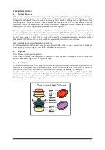
,OOXPLQDWLRQ6\VWHP
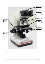
In order to provide sufficient brightness to observe the specimen and make full use of the resolution of the
objectives, this instrument uses a 6V, 20W built-in variable-brightness lamp (see figure 4.0). For optimal results, the
condenser must be adjusted properly. The condenser is composed of two parts. The iris diaphragm is mounted
under the stage, the illumination source is in the base. Adjust the condenser iris (mounted under the stage) to
match the aperture of the objective. The stage can be raised and lowered using the focussing knobs. The light axis
of the condenser must coincide with the light axis of the illumination source. To correct the alignment, adjust 3
screws in the part of the condenser mounted on the stage.
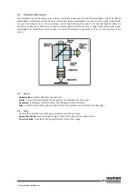
4.2.1
Köhler Illumination
The HUMASCOPE is equipped with Köhler illumination. In order to get the best image possible form the optical
setup of the microscope for brightfield, phase contrast and differential interference contrast, it is crucial that the
light path be set up properly.
HOW TO SET UP HUMASCOPE PROPERLY FOR KÖHLER ILLUMINATION:
If the light path is set up properly, the user has the advantage of an evenly illuminated field, a bright image
without glare and minimum heating of the specimen.
The following instructions apply to a microscope equipped for transmitted-light brightfield illumination..
1. Switch on the light source and make sure that light is passing through the field diaphragm at the base of
the microscope stand. It may help to place a piece of paper over the field stop to confirm this.
Place your specimen on the stage and turn the nosepiece (which holds the objective lenses) to the 10X or
20X lens. Open the field diaphragm as far as possible.
2. Check if the specimen is illuminated. It will help to place a piece of paper over the top of the specimen to see
if light is getting through. If you are using the brightfield condenser stop, open the iris diaphragm (or
aperture diaphragm) on the condenser turret (which contains the stops for brightfield and phase, etc) to the
maximum. The front lens should be about 1-3 mm above the specimen. Use the condenser focussing knobs
to do this.
3. Bring the specimen into focus with the coarse and fine focussing knobs. If the light is too bright, reduce the
light intensity using the light adjustment wheel.
4. When the specimen is in focus, start to close the field diaphragm and also begin to carefully move the
condenser up and down with the condenser focussing knobs. Look for a sharp image of the edge of the field
diaphragm.
5. When the edge of the field diaphragm silhouette is sharply defined, centre it with the two knurled knobs
that extend diagonally from the condenser. Close down the field diaphragm most or all the way to get it
centred properly. When it is centred, open the field diaphragm until its edge is outside the field.
6. Your specimen should now be properly illuminated and you should have a good image.
0HFKDQLFDO6WDROGHU
The HUMASCOPE features a double-layer stage mechanism. This mechanism allows the operator to move the
specimen slide along the X or Y axis by turning the corresponding knob. This is very useful at higher magnifications,
as it can be difficult to move the slide in small enough increments by hand. Also, moving the slide by hand can be
difficult since it must be moved in the opposite direction of the observed movement. The mechanical stage also
has a graduated scale so you can see how far the slide has been moved or keep track of the position of various
objects on the slide.