background image

7/9 

+XPD6FRSH


8VHU0DQXDO

5


 OPERATION

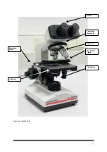
1.  Screw the objectives into the threaded holes in the nosepiece in order of their magnification. Insert the oculars

into the inclined ocular tube.

2.  Place a specimen slide on the stage. Adjust the stage so that the specimen is centred over the hole in the stage.

3.  Using first the 10X objective, turn the coarse focussing knob to move the objective close to the specimen. Now

observe the image through ocular and turn the coarse focussing knob to lower the objective until the image

comes into approximate focus. Next, turn the fine focussing knob until the image is sharp. Thanks to the

instrument’s precise and reliable nosepiece, a clear focus is maintained even when switching objectives.

4.  The specimen can also be observed by replacing the lower condenser with a plan-concave reflecting mirror.

6


STANDARD ACCESSORIES

     ITEMS

CONTENTS

QUANTITY

       1

Ocular WF10X, P16X

2PCS each

       2

Objective 4X 10X 40X 100X

1PC each

       3

Reflecting Mirror

1

       4

Spare Lamp

1

       5

Spare Fuse

1

       6

Immersion Oil

1BOTTLE

       7

User Manual

1

       8

Blue Filter

1

       9

Power Cable

1

     10

Dust Cover HUMAN

1

7


CARE AND MAINTENANCE:

1. As with any other optical instrument, the microscope should be kept in a cool, dry, dust- and acid-free location.

The instrument should be covered with dust cover after use.

2. Never disassemble the lenses; these are precisely adjusted and glued in place at the factory. Stains can be

removed from the lens by wiping it carefully with a soft, clean cloth moistened with alcohol. Take care,

however, that alcohol does not seep it into the objectives to avoid dissolving the glue. Dust on the lens can be

removed using a clean lens brush.

3.  The coarse and fine focussing assembly and nosepiece are precisely adjusted at the factory and should not be

dismantled.

4.  When not in use, keep the objectives in the objective box and cover the ocular tube with the ocular-tube cover.

8


DISPOSAL MANAGEMENT CONCEPT:

The currently valid local regulations governing disposal must be observed. It is in the responsibility of the user to

arrange for proper disposal of the individual components.

All parts which have come into contact with potentially infectious materials must be disinfected using a valid

procedure (autoclaving, chemical treatment) prior to disposal. Applicable local regulations for disposal must be

carefully observed.

The instrument and electronic accessories (without batteries, power packs etc.) must be disposed of according to

regulations for the disposal of electronic components.

Batteries, power packs etc. must be separated from electric/electronic parts and disposed of in accordance with

applicable local regulations.

Содержание HumaScope

Страница 1: ...Gtl Rbnod User Manual Cat No 16100 1...

Страница 2: ...i Qduhrhnm Khrs ne sgd L mt k No DATE Rev REVISION DESCRIPTION 1 01 2004 07 First edition 2 02 2006 08 Addition of Chapter 4 2 1 K hler Illumination Update Specifications...

Страница 3: ...ii...

Страница 4: ...llation of this product This warranty does not apply to damages incurred in shipment of goods Any damage so incurred shall be reported to the freight carrier for settlement or claim 3 INTENDED USE OF...

Страница 5: ...at least potentially infectious Before doing any servicing on the instrument it is very important to thoroughly disinfect all possibly contaminated parts Before the instrument is removed from the labo...

Страница 6: ...Holder 4 4 4 Ocular Objective and Nosepiece 5 4 5 Coarse and Fine Focussing Knobs 5 4 6 Condenser 5 4 7 Immersion Oil 5 5 OPERATION 7 6 STANDARD ACCESSORIES 7 7 CARE AND MAINTENANCE 7 8 DISPOSAL MANA...

Страница 7: ...2 9...

Страница 8: ...trument It can also be used in research work such as biology bacteriology cytology histology and pharmaceutical chemistry 3 SPECIFICATIONS Ocular Designation Magnification Field Of View Diameter Focal...

Страница 9: ...e nosepiece which holds the objective lenses to the 10X or 20X lens Open the field diaphragm as far as possible 2 Check if the specimen is illuminated It will help to place a piece of paper over the t...

Страница 10: ...focus the stopper may be used to lock in the focus This also allows the stage to be lowered and raised again to exactly the same position When the position is no longer needed simply loosen the stopp...

Страница 11: ...6 9 Figure 4 0 HUMASCOPE Light Source Mechanical Stage Revolving Nosepiece Ocular Objective Focussing Knobs Condenser...

Страница 12: ...free location The instrument should be covered with dust cover after use 2 Never disassemble the lenses these are precisely adjusted and glued in place at the factory Stains can be removed from the le...

Страница 13: ...ible Besides the optical advantages darkfield illumination is very beautiful and gives an almost science fiction like image What is the difference between darkfield and brightfield The difference betw...

Страница 14: ...light source with proper wavelengths for excitation is also needed For normal fluorescence applications this is a mercury vapour arc burner HQVHV objective lens gathers light from the specimen ocular...

Страница 15: ...HUMAN Gesellschaft f r Biochemica und Diagnostica mbH Max Planck Ring 21 65205 Wiesbaden Germany Tel 49 61 22 99 88 0 Fax 49 61 22 99 88 100 e Mail human human de www human de 02 2006 08...

Отзывы: