11 |
NexGen MIS LPS-Flex Mobile Implant System
Surgical Technique
MIS Subvastus Approach
Becoming accustomed to operating through a small
incision and adopting the concept of a mobile window
may be facilitated by starting with a shortened medial
parapatellar arthrotomy. This will help to improve
visualization of the anatomy during the initial stages of
becoming familiar with an MIS approach.
When comfortable with the MIS medial parapatellar
approach, performing the arthrotomy through a
midvastus approach will help preserve the quadriceps
tendon and a portion of the medial muscular
attachment. As this procedure becomes more familiar,
the level of the midvastus incision should be lowered
to maintain more muscle attachment.
The subvastus arthrotomy provides excellent exposure
through an MIS incision. The oblique portion of the
incision starts below the vastus medialis obliquus
(VMO) attachment and will preserve all the medial
muscle attachments, including the retinacular
attachment to the medial patella. A key aspect of the
subvastus approach is that it is not necessary to evert
the patella. This helps avoid tearing of the muscle
fibers and helps maintain muscle contraction soon
after surgery.
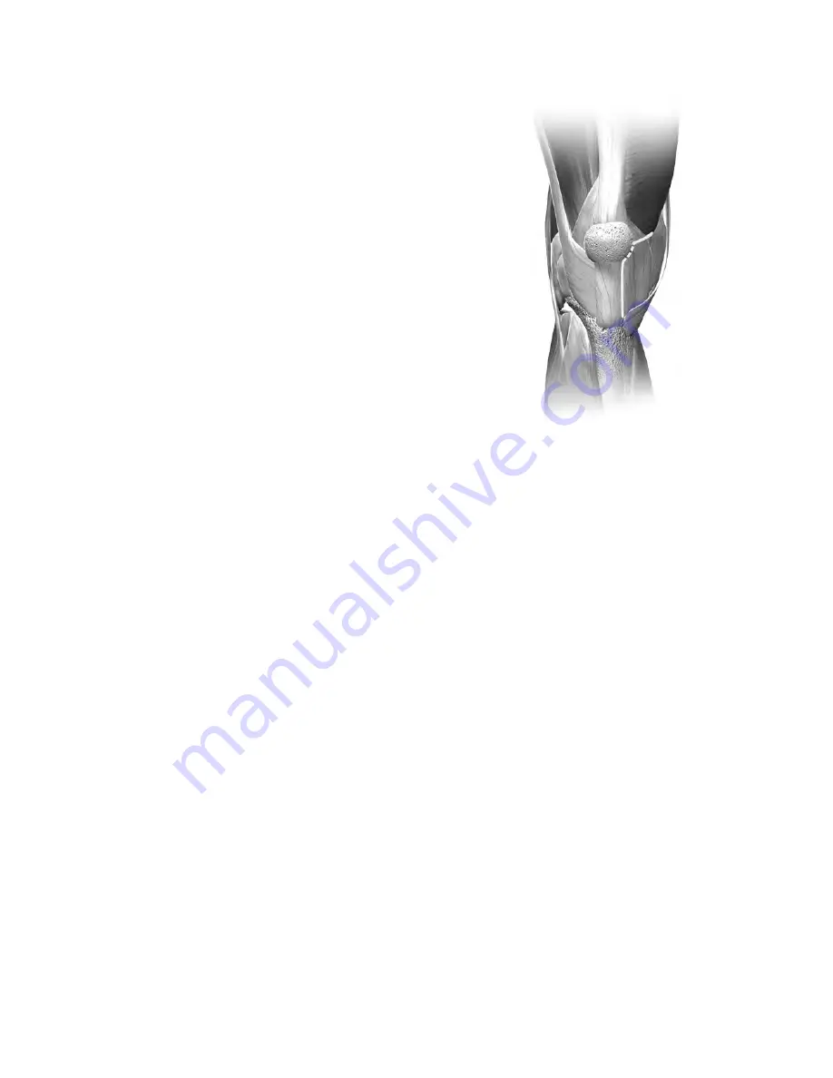
The longitudinal incision should extend only to the
point of insertion of the VMO inferiorly, not to the
proximal pole. Begin the arthrotomy at the medial
edge of the tubercle and extend it along the border
of the retinaculum/tendon to a point on the patella
corresponding to 10 o’clock on a left knee or 2 o’clock
on a right knee. Then continue the incision obliquely
1 cm-2 cm just below and in line with the VMO fibers
(Figure 8). Do not extend the oblique incision beyond
this point as it creates further muscle invasion without
providing additional exposure.
Perform a medial release according to surgeon
judgment, depending on the degree of varus or valgus
deformity. To facilitate a medial release, place the knee
in extension with a rake retractor positioned medially
to provide tension that will assist in developing this
plane. For valgus deformities, consider performing
a more conservative medial release to avoid over-
releasing an already attenuated tissue complex.
With the knee in extension and a rake retractor
positioned to place tension on the patella, remove the
retropatellar fat pad. Then excise a small piece of the
capsule at the junction of the longitudinal and oblique
retinacular incisions. This release allows the patella to
retract laterally. Undermine the suprapatellar fat pad,
but do not excise it. This helps ensure that the femoral
A/P sizer will be placed directly on bone rather than
inadvertently referencing off soft tissue, which may
increase the femoral size measurement.
Placement of a lateral retractor is very important for
adequate retraction of the patella. With the knee
extended, slip the retractor into the lateral gutter and
lever it against the retinaculum at the superomedial
border of the patella. As the knee is flexed, the patella
is retracted laterally to provide good visualization of
the joint.
Figure 8


