


PD110323 Rev. A
1
MicroVention, Inc.
English
Azur
®
CX Peripheral Coil System
(Detachable)
Instructions for Use
DEVICE DESCRIPTION
The Detachable Azur CX Peripheral Coil System (Azur system) consists of a coil
implant attached to a delivery system. The coils are platinum coils with an inner
layer of hydrophilic polymer. The delivery pusher is powered by an Azur Detachment
Controller to selectively detach the coils. The Azur Detachment Controller is provided
separately.
The Azur system is available in a broad range of coil diameters and lengths. The coil
must be delivered only through a wire-reinforced microcatheter with the inner diameter
specified.
Table 1
Coil Type
Microcatheter I.D.
Reposition
Time
inches
mm
Azur Detachable 18
0.021 - 0.027
0.53 - 0.69
30 minutes
INDICATIONS FOR USE
The Azur system is intended to reduce or block the rate of blood flow in vessels
of the peripheral vasculature. It is intended for use in the interventional radiologic
management of arteriovenous malformations, arteriovenous fistulae, aneurysms, and
other lesions of the peripheral vasculature.
This device should only be used by physicians who have undergone training in the use
of the Azur system for embolization procedures as prescribed by a representative from
Terumo or a Terumo-authorized distributor.
CONTRAINDICATIONS
Use of the Azur system is contraindicated in any of the following circumstances:
• When superselective coil placement is not possible.
• When end arteries lead directly to nerves.
• When arteries supplying the lesion to be treated are not large enough to accept
emboli.
• When the A-V shunt is larger than the coil.
• In the presence of severe atheromatous disease.
• In the presence of vasospasm (or likely onset of vasospasm).
POTENTIAL COMPLICATIONS
Potential complications include, but are not limited to: hematoma at the site of entry,
vessel/aneurysm perforation, unintended parent artery occlusion, incomplete filling,
vascular thrombosis, hemorrhage, ischemia, vasospasm, edema, coil migration or
misplacement, premature or difficult coil detachment, clot formation, revascularization,
post-embolization syndrome, and neurological deficits including stroke and possibly
death.
The physician should be aware of these complications and instruct patients when
indicated. Appropriate patient management should be considered.
REQUIRED ADDITIONAL ITEMS
• Azur Detachment Controller
• Wire-reinforced microcatheter with distal tip RO marker, appropriately sized
• Guide catheter compatible with microcatheter
• Steerable guidewires compatible with microcatheter
• 2 rotating hemostatic Y valves (RHV)
• 1 three-way stopcock
• Pressurized sterile saline drip
• 1 one-way stopcock
• Stopwatch or timer
WARNINGS AND PRECAUTIONS
Caution: Federal law (USA) restricts this device
to sale by or on the order of a physician.
• The Azur system is supplied sterile and non-pyrogenic unless package is opened
or damaged.
• This device is intended for single use only. Do not reuse, reprocess or resterilize.
Reuse, reprocessing or resterilization may compromise the structural integrity of
the device and/or lead to device failure which, in turn, may result in patient injury,
illness, or death. Reuse, reprocessing, or resterilization may also create a risk of
contamination of the device and/or cause patient infection or cross-infection,
including, but not limited to, the transmission of infectious disease(s) from one
patient to another. Contamination of the device may lead to injury, illness or death
of the patient.
• Angiography is required for pre-embolization evaluation, operative control, and
post-embolization follow up.
• Do not advance the delivery pusher with excessive force. Determine the cause of
any unusual resistance, remove the Azur system, and check for damage.
• Advance and retract the Azur system slowly and smoothly. Remove the entire Azur
system if excessive friction is noted. If excessive friction is noted with a second
Azur system, check the microcatheter for damage or kinking.
• The coil must be properly positioned in the vessel or aneurysm within the specified
reposition time from the time the device is first introduced into the microcatheter.
If the coil cannot be positioned and detached within this time, simultaneously
remove the device and the microcatheter. Positioning the device in a low-flow
environment may increase the reposition time.
• If repositioning is necessary, take special care to retract the coil under fluoroscopy
in a one-to-one motion with the delivery pusher. If the coil does not move in a one-
to-one motion with the delivery pusher, or if repositioning is difficult, the coil may
have become stretched and could possibly break. Gently remove and discard the
entire device.
• Due to the delicate nature of the coils, the tortuous vascular pathways that lead
to certain lesions, and the varying morphologies of the vasculature, a coil may
occasionally stretch while being maneuvered. Stretching is a precursor to potential
coil breakage and migration.
• If a coil must be retrieved from the vasculature after detachment, do not attempt to
withdraw the coil with a retrieval device, such as a snare, into the delivery catheter.
This could damage the coil and result in device separation. Remove the coil,
microcatheter, and any retrieval device from the vasculature simultaneously.
• Delivery of multiple coils is usually required to achieve the desired occlusion
of some vasculatures or lesions. The desired procedural endpoint is usually
angiographic occlusion. The filling properties of the coils facilitate angiographic
occlusion.
• Tortuosity or complex vessel anatomy may affect accurate placement of the coil.
• The long-term effect of this product on extravascular tissues has not been
established so care should be taken to retain this device in the intravascular space.
• Always ensure that at least two Azur Detachment Controllers are available before
starting an Azur system procedure.
• The coil cannot be detached with any power source other than an Azur
Detachment Controller.
• Do NOT place the delivery pusher on a bare metallic surface.
• Always handle the delivery pusher with surgical gloves.
• Do NOT use in conjunction with radio frequency (RF) devices.
PREPARATION FOR USE
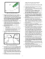
1.
Refer to Figure 1 for the set-up diagram.
2.
Attach a rotating hemostatic valve (RHV) to the hub of the guiding catheter.
Attach a 3-way stopcock to the side arm of the RHV and then connect a line for
continuous infusion of flush solution.
3.
Attach a second RHV to the hub of the microcatheter. Attach a 1-way stopcock
to the sidearm of the second RHV and connect the flush solution line to the
stopcock.
4.
Open the stopcock and flush the microcatheter with sterile flush solution and
then close the stopcock. To minimize the risk of thromboembolic complications,
it is critical that a continuous infusion of appropriate sterile flush solution be
maintained into the guide catheter, the femoral sheath and the microcatheter.
CATHETERIZATION OF THE LESION
5.
Using standard interventional procedures, access the vessel with a guide
catheter. The guide catheter should have an inner diameter (ID) large enough to
allow for contrast injection while the microcatheter is in place. This will allow for
fluoroscopic road mapping during the procedure.
6.
Select a microcatheter with the appropriate inner diameter. After the
microcatheter has been positioned inside the lesion, remove the guidewire.
COIL SIZE SELECTION
7.
Perform fluoroscopic road mapping.
8.
Measure and estimate the size of the lesion to be treated.
9.
For aneurysm occlusion, the diameter of the first and second coils should never
be less than the width of the aneurysm neck or the propensity for the coils to
migrate may be increased.
10.
For vessel occlusion, select a coil size that is slightly larger than the vessel
diameter.
11. Correct coil selection increases effectiveness and patient safety. Occlusive
efficiency is, in part, a function of compaction and overall coil mass. In order
to choose the optimum coil for any given lesion, examine the pre-treatment
angiograms. The appropriate coil size should be chosen based upon
angiographic assessment of the diameter of the target or parent vessel,
aneurysm dome and aneurysm neck.


